Case Report: 5 Years Old Boy with Skin Morphea Appeared in the Same Site of a Previous Chronic Eczematous Lesion. King Abdulaziz Medical City of the National Guard. Saudi Arabia-September 2019.
Case Report: 5 Years Old Boy with Skin Morphea Appeared in the Same Site of a Previous Chronic Eczematous Lesion.
King Abdulaziz Medical City of the National Guard. Saudi Arabia-September 2019.
Dr. Ahmed Moosa Yahya assery*
*Correspondence to: Dr. Ahmed Moosa Yahya assery, Paediatrics ambulatory care consultant. King Abdulaziz Medical City, National Guard Medical Affairs, Family Medicine-Ambulatory care, NGCSC.
Copyright© 2019: Dr. Ahmed Moosa Yahya assery. This is an open access article distributed under the Creative Commons Attribution License, which permits unrestricted use, distribution, and reproduction in any medium, provided the original work is properly cited.
Received: 06 November 2019
Published: 15 November 2019
Abstract
Morphia (mor-FEE-uh) is a rare condition that causes painless, discoloured patches on your skin. typically, the skin changes appear on the belly, chest or back. But they might also appear on your face, arms and legs. Over time the patches may become firm, dry and smooth. Morphea tends to affect only the outer layers of your skin. But some forms of the condition also affect deeper tissues and may restrict movement in the joints. Morphea usually improves on its own over time, though recurrences are common. In the meantime, medications and therapies are available to help treat the skin discoloration and other effects.in our reported case her the striking thing is the appearance of the morphia lesion in the site of the body (lower neck) that presented with lesion of atopic dermatitis ( Eczema) for long time which triggered our curiosity to ask ourself ,is there any histopathological relation between the two entities of dermatological disorder (morphea and eczema) or not? the key that may open for us the gate to solve the secrets of the morphea in the pathogenesis and the treatment wise.(1).
Keywords: KingAbdullAziz Medical City: KAMC.
National Guard Comprehensive Specialized Clinics: NGCSC.
Morphea: MPH.
juvenile localized scleroderma: JLS.
Juvenile systemic sclerosis: JSSc.
systemic sclerosis: SSc
Case Report: 5 Years Old Boy with Skin Morphea Appeared in the Same Site of a Previous Chronic Eczematous Lesion. King Abdulaziz Medical City of the National Guard. Saudi Arabia-September 2019.
Introduction
(Morphea): Although scleroderma is a spectrum of disorders that can occur at any stage of life, the clinical patterns of childhood scleroderma differ from that of adulthood. The predominant form of scleroderma in children is juvenile localized scleroderma (JLS), also sometimes called MPH, which principally involves the skin, fascia, muscle, and bone. JSSc is a chronic multisystem connective tissue disorder characterized by hardening of the skin accompanied by abnormalities of the visceral organs. JLS is only very rarely associated with systemic disease. However, it can sometimes mimic eosinophilic fasciitis.
Table 1: Classification of the JLS
Classification
JLS is differentiated from SSc based upon the pattern of cutaneous and extracutaneous involvement. In JLS, there is cutaneous involvement, typically with some involvement of the underlying musculature and/or bony structures, but rarely the nervous system, and absence of the internal organ involvement that typifies SSc (i.e., the lungs and gastrointestinal tract).
Various classification systems for JLS MPH subtypes have been proposed, but the most in use are the 1995 Mayo Clinic criteria, the 2006 Padua criteria, and the 2017 European Dermatology Forum criteria. A cross-sectional study in two large patient cohorts compared the performance of these three classifications and found that the Padua criteria performed best in classifying patients into subgroups, supporting its widespread use. The Padua classification criteria divides JLS into five types (table 1) :
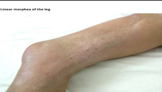
?Linear scleroderma (65 percent) – This is the most common type of JLS in children. The fibrotic lesion presents in a linear distribution, and the orientation is usually transverse when on the trunk and longitudinal on the limbs (picture 1). During the active phase, the border of the lesion is reddish (hyperaemic) while the centre is ivory colour and waxy. During the inactive phase, it can be either hypo- or hyperpigmented. Patients may develop contractures and defects of the limb with associated poor growth and disabilities.
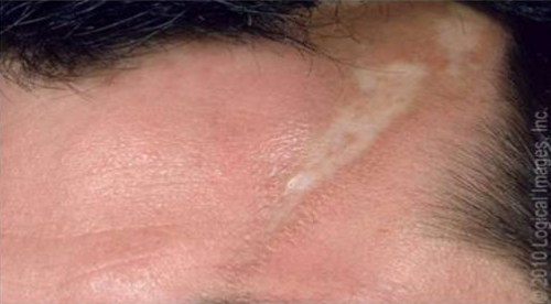
When the lesion involves the face or scalp, it is also referred to as "en coup de sabre" (picture 2).
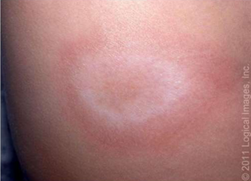
?Circumscribed (plaque) MPH (26 percent) – This is the most benign form of JLS. Circumscribed MPH is characterized by oval or round circumscribed areas of induration with a central waxy, ivory colour surrounded by a violaceous halo. In the superficial variety, the lesion is confined to the dermis with only occasional involvement of the superficial panniculus (picture 3).
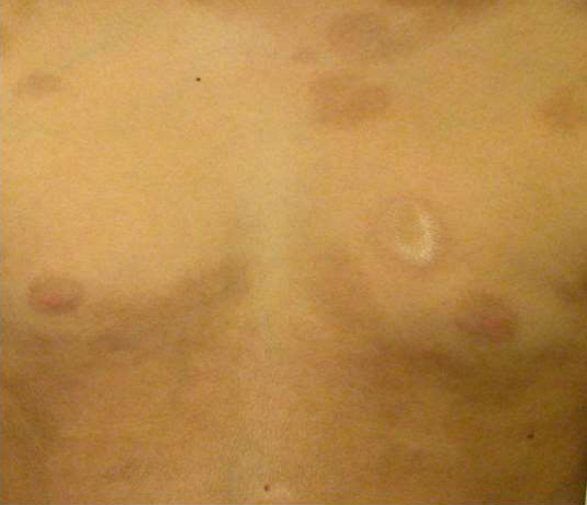
?Generalized MPH (7 percent) – This form of JLS occurs when there are four or more plaques that affect two or more anatomic sites and become confluent (picture 4). Generalized MPH can be superficial or deep.
?Pan sclerotic (deep) MPH (2 percent) – This is the least common but the most disabling form of JLS. The primary site of involvement is the panniculus or subcutaneous tissue.
?Mixed MPH – A combination of two or more of the above subtypes occurs in approximately 15 percent of patients. This overlap occurs most frequently with circumscribed MPH and linear scleroderma.
Picture 1: Linear MPH
Picture 2: secondary MPH
Picture 3: circumscribed MPH
Picture 4: generalized MPH
Epidemiology
Although JLS is uncommon, it is 6 to 10 times more common in children than systemic sclerosis (SSc). Few studies have addressed the incidence or prevalence of this disorder in children. Approximately one-third of all patients with localized scleroderma have disease onset in childhood, with an estimated incidence rate of 0.34 to 0.9 cases per 100,000 children per year age ≤16 years. However, JLS may be underreported because of referral bias patterns and misdiagnosis as other cutaneous disease.
Etiology and pathogenesis
The aetiology and pathogenesis of JLS remain uncertain. Numerous etiologic agents have been suggested as possible causes of JLS, including drugs and environmental toxins, local trauma, and infection. However, no definitive link has been established in any of these. Two possible contributing pathogenic processes include abnormal fibroblast function and immune dysfunction resulting in autoimmunity.
Clinical manifestations
Clinical manifestations vary depending upon the type of JLS.
Age of presentation — The mean age of presentation is 7 to 8.2 years (range: birth to 17 years; median age: 6 years).
Distribution of lesions — In comparison with adults, the head is preferentially affected by linear scleroderma, whereas the trunk is more likely affected by plaque and generalized MPH in children. The frequency of extracutaneous involvement also varies with the location of the lesions. In a retrospective study, patients with lesions on the anterior or superior aspect of the head had a higher risk of neurologic involvement than those with lesions on the back or inferior aspect of the head, and those with extensor extremity lesions were at increased risk of musculoskeletal complications than those with flexor involvement.
Linear scleroderma — Linear scleroderma is the most frequent form of JLS in children The fibrotic lesion appears as linear bands. Limbs are more commonly affected than the face. The orientation of the fibrotic band is usually transverse on the trunk and longitudinal in the extremities. Recurrence in JLS occurs in almost one-quarter of patients and is more frequent in the linear subtype involving the limbs independently of the age of the disease onset. Extracutaneous manifestations are most common in patients with linear scleroderma.
There is considerable debate on whether these lesions follow specific dermatomes. One theory speculates that the involved distribution reflects an abnormal migration of neuroectodermal cells during embryogenesis. This theory is supported by the frequent development of seizures, headaches, and muscle weakness on the affected side during the evolution of facial lesions. Imaging studies of the brain also have shown vascular and cerebral changes even in patients without neurologic symptoms. Other data suggest that linear lesions follow the lines of Blaschko. (2).
Atopic dermatitis: Atopic dermatitis is a chronic, pruritic, inflammatory skin disease that occurs most frequently in children but also affects adults. Atopic dermatitis is often associated with an elevated serum level of immunoglobulin E (IgE) and a personal or family history of atopy, which describes a group of disorders that includes eczema, asthma, and allergic rhinitis. Although sensitization to environmental or food allergens is clearly associated with the atopic dermatitis phenotype, it does not seem to be a causative factor but may be a contributory factor in a subgroup of patients with severe disease.
Epidemiology
Prevalence and incidence — Atopic dermatitis affects approximately 5 to over 20 percent of children worldwide, with large variations among countries and ethnic groups. Countries in Africa, Oceania, and the Asia-Pacific region have higher rates of atopic dermatitis than countries in the Indian subcontinent and Northern/Eastern Europe. In the United States, the overall prevalence is approximately 16 percent, with the highest rates reported in African American children (19 percent).
Data on the prevalence of atopic dermatitis in adults are limited. Population-based studies from Scandinavian countries report prevalence rates of 10 to 14 percent among adults. In a United States, cross-sectional study including nearly 1300 adults, the prevalence of atopic dermatitis was 7.3 percent (95% CI 5.9-8.8).
While the incidence remains high in urban areas and high-income countries, an increasing trend in incidence and prevalence of atopic eczema has been reported in the last few decades in Africa, East Asia, Western Europe, as well as in parts of Northern Europe.
In the vast majority of cases, atopic dermatitis has an onset before the age of five years, and prevalence data in children show a slight female preponderance. Persistent atopic dermatitis beyond infancy may affect approximately 50 percent of patients diagnosed with atopic dermatitis during childhood. Onset in the first six months of life appears to be associated with severe disease.
Risk factors — Risk factors for atopic dermatitis include multiple genetic and environmental factors.
?Genetic risk factors – A family history of atopy (eczema, asthma, or allergic rhinitis) is the strongest risk factor for atopic dermatitis. Approximately 70 percent of patients have a positive family history of atopic diseases. Children with one atopic parent have a two- to threefold increased risk of developing atopic dermatitis, and the risk increases to three- to fivefold if both parents are atopic.
Loss-of-function variants in the FLG gene, resulting in defective epidermal barrier, are a major risk factor for atopic dermatitis and other skin and allergic diseases, including allergic contact dermatitis, asthma, and food allergy. Multiple other genes have been proposed as potential contributors to the risk of atopic dermatitis, including genes involved in the regulation of innate host defences and T cell function.
?Environmental exposures – Environmental factors, including climate, urban versus rural setting, air pollution, early exposure to nonpathogen microorganisms, and water hardness, may influence the risk of atopic dermatitis. Examples of studies linking environmental factors to atopic dermatitis are shown below:
- The "hygiene hypothesis" – Two systematic reviews provided evidence to support an inverse relationship between atopic dermatitis and exposure to endotoxin, early daycare, helminth infestation, number of siblings, farm animals, and pet dogs in early life. There was no protective effect associated with viral or bacterial infections.
- Water hardness – Epidemiologic evidence from ecologic studies linked high hardness (high levels of calcium carbonate) of domestic water with increased prevalence of atopic dermatitis in children. A 2021 meta-analysis of seven observational studies that included nearly 386,000 participants found a modest increase of risk of atopic dermatitis in children exposed to hard water (odds ratio [OR] 1.28, 95% CI 1.09-1.50). However, the authors considered the certainty of this estimate to be very low, due to high risk of bias and heterogeneity in the definition of "hard water."
Pathophysiology
A multiplicity of mechanisms are involved in the pathogenesis of atopic dermatitis, including epidermal barrier dysfunction, genetic factors, Th2 cell-skewed immune dysregulation, altered skin microbiome, and environmental triggers of inflammation. Whether skin inflammation is initiated by skin barrier dysfunction ("outside-in" hypothesis) or by immune dysregulation ("inside-out" hypothesis) is still debated. It is increasingly recognized that combinations of different mechanisms result in multiple "endotypes" and phenotypes of atopic dermatitis.
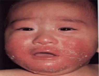
Picture 5: Infantile atopic dermatitis.
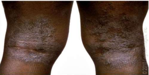
Picture 6: Lichenification of atopic dermatitis
Clinical Manifestation
Common features — Dry skin and severe pruritus are the cardinal signs of atopic dermatitis. However, the clinical presentation is highly variable, depending upon the patient's age, ethnicity, and disease activity.
Acute eczema is characterized by intensely pruritic, erythematous papules and vesicles with exudation and crusting (picture 5), whereas subacute or chronic lesions present as dry, scaly, or excoriated, erythematous papules . Skin thickening from chronic scratching (lithification) and fissuring may develop over time (picture 6). In many patients, lesions in different stages may be present at the same time.
In children and adults with deeply pigmented skin, erythema may appear dark brown or violaceous instead of pink or red, as typically seen in patients with lighter complexions. The typical erythematous and scaly lesions of eczema may appear as lesions with a greyish, violaceous, or dark brown hue. Dry skin may have a whitish or ashy colour and a reduction in skin shininess (Lichenified areas typically appear hyperpigmented. Post inflammatory hyper- and hypopigmentation are also common.
Atopic dermatitis occurs in the first year of life in 60 percent of cases and by the age of five years in nearly 85 percent of cases. The clinical presentation at various ages is outlined below:
?In infants and young children (zero to two years), atopic dermatitis typically presents with pruritic, red, scaly, and crusted lesions on the extensor surfaces and cheeks or scalp but may be diffuse. There is usually sparing of the diaper area. Acute lesions can include vesicles, and there can be serous exudates and crusting in severe cases.
?In older children and adolescents (2 to 16 years), atopic dermatitis is characterized by less exudation and often demonstrates lichenified plaques in a flexural distribution, especially of the antecubital and popliteal fossae, volar aspect of the wrists, ankles, and neck. The sides of the neck may show a reticulate pigmentation, the so-called "atopic dirty neck. (3).
Our Case Presentation
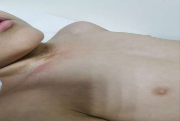
This child who was 5 years when presented to my clinic for the first time the mother concern was about a strange skin lesion that started to grow gradually in his lower neck ( at the junction with the upper chest) over a period of 2 months (picture 7 )and according to the mother that lesion was a site of long time scaly erythematous itchy lesion that was reoccurring and disappearing in an on-off manner who since birth , that red old skin manifestation seen by a dermatologist ( according to the mother ) and labeled as an eczema which was responding very dramatically to the topical moisturizer and hydrocortisones, the mother among the counselling in my clinic she approved the presence of significant family history of eczema and denied any other family history of psoriasis or morphea.
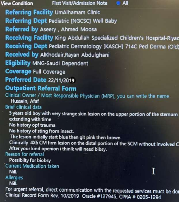
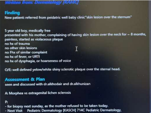
Soon after the visit and according to our medical institution authorized organizations I referred the case to the pediatrics dermatology (picture 8) who saw the patient and labeled him as a case of query morphea VS extragenital lichen sclerosis and decided to proceed to the skin biopsy as soon as possible (picture 9).
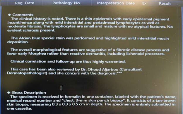
The biopsy done and it was strongly suggestive of early morphea (picture 10) then the family has been informed about the result and reassured about the diagnosis and the modality of treatment (mid-potent topical hydrocortisone).
A follow up planed with dermatology clinic after the discharge and the family informed about the possible psychological sequalae that may happened later in form of self-steamed issues other than that no major and dangerous complication will be suspected.
Picture 7: The skin lesion that our case presented with.
Picture 8: The case referral request from me to the dermatologist
Picture 9: The first visit note written by the dermatologist
Picture 10: The final biopsy report of the case written by the histopathologist
Discussion about the Case
The interesting tow point in this case that I want to high light is as follow:
1) The discovery of such rare dermatological disorder (MPH) in our institution where as we mentioned previously that epidemiologically the estimated incidence rate of 0.34 to 0.9 cases per 100,000 children per year age ≤16 years. However, JLS may be underreported because of referral bias patterns and misdiagnosis as other cutaneous disease. (2).
In other site in the (4) in American journal of dermatology ( published in 2017 , Volume 18,pages 491-512. March 2017 by • Jorre S. Mertens, • Marieke M. B. Seyger, • Rogier M. Thurlings, • Timothy R. D. J. Radstake & • Elke M. G. J. de Jong ) it was mentioned that The rarity of MPH is reflected in the annual incidence rates which are reported to be between 3.4 and 27 cases per1,000,000 . Females are more frequently affected than males (ratio: 2.4–5.0 to 1). The peak incidence is bimodal with peaks between 7 and 11 years for paediatric onset disease and 44–47 years for adult-onset disease. The incidence and prevalence of EF is unknown. The disease predominantly affects patients in their fourth and fifth decade of life. Only a few case reports describe childhood-onset disease.
See also in this article (5) Journal of the American Academy of Dermatology ( Volume 80, Issue 6, June 2019, Pages 1664-1670.e1 Elaine Kunzler MD a b, Stephanie Florez Pollack BS a, Noelle Teske MD,MSc a, Jack O'Brien MD a, Smriti Prasad BS a, Heidi Jacobe MD, MSC) it mentioned that Linear MPH was the most common MPH subtype (50.1%, 291/581) in the cohort. Deep involvement was more common in linear (64.3%, 187/291) than other MPH subtypes. Linear MPH participants with deep involvement were more likely to have a limitation in range of motion (28.6%, 55/192) than those without (11.1%, 11/99, P < .001). Adult-onset disease occurred in 32.6% (95/291) of those with linear MPH. Frequency of deep involvement was similar between paediatric (66.8%, 131/196) and adult-onset linear MPH (58.9%, 56/95, P = .19). Quality of life and disease activity scores improved over time, while damage stabilized with treatment.
So, from the first point we should agree all about the rarity of MPH, which makes our article so expensive and important for further research into it in order to discover many secrets behind this rare disease.
2) Then incidental occurrence between the morphea and the eczema (as in our case report) that deserve further investigations and elaboration, from the Medline search nothing at all joined the two entities in any way except one article (6) was taking about New Light Therapy Effective Treatment for MPH, Eczema (mentioned by UT Southwestern medical centre in 28-Jan-2004 6:50 AM EST) that mentioned Phototherapy is beneficial because it can target large areas of skin without the side effects of oral or topical medications. UVA-1 is used to treat some patients with inflamed skin, or eczema, especially those with hand dermatitis or dyshidrosis " a condition that causes blisters on the palms of the hands and soles of the feet " and MPH, thought to be an autoimmune disease that is localized to the skin and causes thickening and discoloration.
Being treating both of MPH and eczema by the same manner and steps could be a big clue that both of them my originate of the same stem of pathology.
So, the question is this rare, mysterious morphea disorder triggered by a preexisting clinical or subclinical Eczema or not?
Conclusion
Morphea is still a rare dermatological disorder not only in adults but rather in the pediatric group of people, Eczema may be one of the triggering factors of its occurrence.
References
1-https://www.mayoclinic.org/diseases-conditions/morphea/symptoms-causes/syc-20375283#:~:text=Morphea%20(mor%2DFEE%2Duh,become%20firm%2C%20dry%20and%20smooth.
2_ https://www.uptodate.com/contents/juvenile-localized-scleroderma?source=history
3_ https://www.uptodate.com/contents/atopic-dermatitis-eczema-pathogenesis-clinical-manifestations-and-diagnosis?source=history_widget
4_ https://www.ncbi.nlm.nih.gov/pmc/articles/PMC5506513/
5_ https://pubmed.ncbi.nlm.nih.gov/31005342/
6_https://www.newswise.com/articles/new-light-therapy-effective-treatment-for-morphea-eczema.