The Significance of Erythrocyte Rigidity in the Development of Microangiopathy in Patients with Type 2 Diabetes Mellitus.
The Significance of Erythrocyte Rigidity in the Development of Microangiopathy in Patients with Type 2 Diabetes Mellitus.
Rasheed Manasrah*, Valentin Smorzhevskyi1.
1. Doctor of Medical Sciences, Professor of the Department of Surgery and Transplantology. Shupyk National Healthcare University of Ukraine- Kyiv., National Institute of Surgery and Transplantology named after O. O. Shalimov.
*Correspondence to: Rasheed Manasrah, PhD student of the Department of Surgery and Transplantology, Shupyk National Healthcare University of Ukraine- Kyiv., National Institute of Surgery and Transplantology named after O. O. Shalimov.
Copyright
© 2024: Rasheed Manasrah. This is an open access article distributed under the Creative Commons Attribution License, which permits unrestricted use, distribution, and reproduction in any medium, provided the original work is properly cited.
Received: 20 Sept 2024
Published: 27 Oct 2024
The Significance of Erythrocyte Rigidity in the Development of Microangiopathy in Patients with Type 2 Diabetes Mellitus.
Introduction
Rheological parameters (from the Greek "rheos" – flow) are properties of blood related to its flowability. In large vessels, inertial forces related to mass contribute most significantly to blood flow. However, in the microvascular system, the movement of blood flow is primarily influenced by its rheological properties. This is due to the higher resistance to blood flow in this area, which is determined by the architecture of the vascular system. The rheological properties of blood largely depend on blood viscosity – an integral parameter that defines blood flow. Key factors affecting the viscosity of whole blood include the volumetric concentration of formed elements in plasma, plasma properties, and cellular components' aggregation and deformability.
Since the number of erythrocytes in the bloodstream far exceeds that of other formed elements (accounting for 93–98% of the total), the rheological properties, including blood viscosity, are predominantly determined by the volumetric concentration of erythrocytes. Thus, blood viscosity depends on the hematocrit level, plasma viscosity, and the ability of erythrocytes to aggregate and deform.
Early disability and mortality in patients with diabetes mellitus (DM), typically caused by diabetic angiopathies, represent a significant medical and social issue in contemporary diabetology. Timely detection of microcirculatory disorders and correction of hemorheological abnormalities are now recognized as crucial components of modern diagnostic and therapeutic strategies for patients with diabetes. The micro-rheological properties of erythrocytes, such as deformability, aggregation capacity, and the generation of vasoactive factors, are determined by the characteristics of the molecular organization of the erythrocyte membrane and the perimembrane cytoplasmic matrix.
The polarization of cells, along with the levels of hemolysis and aggregation, is associated with altered conditions during the maturation processes of red blood cells, demonstrating their reduced resilience to various factors. This, in turn, affects their interaction with altered endothelial cells.
• Thus, the rigidity of erythrocytes, measured using atomic force microscopy (AFM), provides valuable information directly related to the development of rheological disturbances in diabetes mellitus.
• Our research indicates that atomic force microscopy may be a promising tool for detecting microcirculatory disturbances in type 2 diabetes.
Keywords:
• Rheology
• Diabetes mellitus
• Erythrocytes
• Rigidity
• Atomic force microscopy (AFM)
Objective of the study:
• To present the diagnostic capabilities for detecting rheological disturbances in patients with type 2 diabetes, based on the measurement of erythrocyte rigidity and viscoelastic parameters using AFM.
Materials and Methods
Twelve patients with type 2 diabetes mellitus (mean age 58.7 ± 1.6 years) were examined, including 3 (25%) women and 9 (75%) men. The control group consisted of 4 subjects with a mean age of 48.5 ± 2.2 years—2 (50%) women and 2 (50%) men—who did not have diabetes or any other manifest pathology of internal organs and presented for preventive purposes (Group 1).
All patients underwent a comprehensive clinical and instrumental examination, including ECG, ultrasound of the abdominal organs, kidneys, heart, and blood vessels, as well as assessments of glycated hemoglobin and the albumin-to-creatinine ratio in a spot urine sample.
Atomic force microscopy (AFM) is an excellent tool for studying altered erythrocytes in patients with type 2 diabetes. Morphological changes in erythrocytes can lead to vascular complications, particularly peripheral arterial disease (PAD), which is a serious complication for individuals living with this condition.
Additionally, it serves as an important indicator of the effectiveness of various surgical interventions and optimal pharmacological therapy in patients with type 2 diabetes.
The viscoelastic parameters of erythrocytes were investigated using atomic force microscopy (AFM). Principle of Atomic Force Microscopy (AFM) Operation.
Results and Discussion
According to the criteria for assessing the risk of vascular complications in patients with type 2 diabetes mellitus (T2DM) proposed by the European Association for the Study of Diabetes (EASD, 2013), patients with T2DM were divided into two groups: those with moderate risk of microvascular damage (moderate degree of rheological – microcirculatory disturbances) – Group 2 (n=7) and those with high risk of microcirculatory disturbances – Group 3 (n=5). The indicators of patients in these groups fell within the values corresponding to the risk of large and small vessel damage.
Data from the clinical and instrumental examinations of the groups are presented. No significant differences were found in age, gender, alcohol consumption patterns, or smoking among patients with T2DM. However, in the group with a high degree of rheological disturbances (Group 3), the duration of diabetes, the length of hypertension, and the frequency of a family history of early cardiovascular diseases were significantly higher. Additionally, there was a higher prevalence of coronary artery disease with a history of myocardial infarctions in this group.
Patients in Group 3 had significantly higher systolic and diastolic blood pressure readings compared to Group 2, with more frequent occurrences of arrhythmias and conduction disturbances (p=0.048). All patients with T2DM received hypoglycemic therapy aimed at achieving target parameters for carbohydrate metabolism control, as well as antihypertensive medications.
Patients with a high degree of rheological disturbances exhibited more severe manifestations of type 2 diabetes mellitus (T2DM) and its complications. This group showed higher levels of fasting blood glucose (9.62 ± 0.69 vs. 7.94 ± 0.33 in Group 2, p = 0.018), glycated hemoglobin, albumin-to-creatinine ratio in a spot urine sample, and more significant signs of hyperlipidemia (predominantly type 2B), as well as disturbances in purine metabolism, liver function, and renal excretory function.
It is noteworthy that patients with pronounced rheological disturbances had a body mass index (BMI) between 35 and 45, greater severity of diabetic neuropathy and retinopathy (with proliferative retinopathy identified in 60% of patients), and angiopathy (in 53.3% of cases, it was classified as stage IIb, where pain in the lower extremities occurred after walking less than 200 meters).
The analysis of red blood cell parameters did not reveal significant differences between the groups in terms of the number of erythrocytes, mean corpuscular volume, or hemoglobin levels. However, it was found that patients with type 2 diabetes mellitus (T2DM) had increased hematocrit and greater width of erythrocyte distribution by volume, while mean corpuscular volume and mean hemoglobin content per erythrocyte were lower than those of healthy individuals.
It is known that the deformability of red blood cells is determined by their viscoelastic characteristics. The results of this study indicated a reduction in the ability of erythrocytes to deform, coinciding with an increase in generalized viscosity and rigidity parameters of the cells. These changes may be associated with an elevated cholesterol content in the erythrocyte membrane and an increased cholesterol-to-phospholipid ratio.
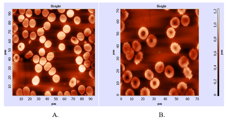
Figure 1
Erythrocytes under AFM: A– in healthy individuals, B– in patients with type 2 diabetes mellitus.
• In comparison to healthy cells, erythrocytes from individuals with type 2 diabetes mellitus (T2DM) exhibited several morphological changes. Specifically, these cells showed a reduced concave depth, diameter, height, and deformation index. Conversely, parameters such as axial ratio, stiffness, adhesive force, aggregation, and rigidity index were found to be increased. The findings related to erythrocyte roughness, however, remained inconclusive.
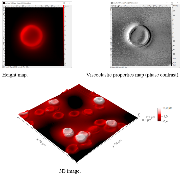
Figure 2
Measurements were conducted using a scanning probe microscope NanoScope IIIa Dimension 3000 (Bruker Inc., formerly Digital Instruments, USA) in intermittent contact mode with a silicon probe having a nominal tip radius of 10 nm.
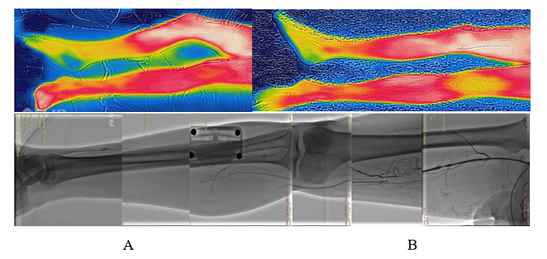
Figure 3, Figure 4
Thermographic clinical assessment of microcirculation disturbances in a patient with diabetes mellitus: photo (A) before treatment, and photo (B) three months after treatment using cell therapy and endovascular angioplasty of the tibial arteries.
Conclusion
- The data obtained indicate that extensive areas of the capillary network become "shut down" from blood flow and oxygen exchange, as aggregates and rigid erythrocytes are unable to pass through the capillaries.
- The study of erythrocyte rigidity using atomic force microscopy (AFM) provides information about erythrocyte deformation and its significance in the development of microangiopathy in type 2 diabetes mellitus.
- Further investigation of erythrocyte rigidity may help in selecting optimal pharmacological treatments for this condition.
References
1. Baskurt, O. K., & Meiselman, H. J. (2003). Blood rheology and hemodynamics. *Seminars in Thrombosis and Hemostasis*, 29(5), 435–450. https://doi.org/10.1055/s-2003-44551
2. Vaya, A., Martinez Triguero, M. L., Alis, R., & Hernandez-Mijares, A. (2010). Erythrocyte deformability in diabetes and association with kidney damage. *Diabetes/Metabolism Research and Reviews*, 26(8), 645–651. https://doi.org/10.1002/dmrr.1137
3. Schmid-Schönbein, H. (1987). Biomechanics of microcirculation: erythrocyte and leukocyte deformability and its implications in diabetes mellitus. *Journal of Biomechanics*, 20(9), 899–909. https://doi.org/10.1016/0021-9290(87)90321-6
4. Tripolino, C., Irace, C., Gnasso, A., & Scavelli, F. (2017). Hemorheological alterations in diabetes mellitus: A state-of-the-art review. *Frontiers in Physiology*, 8, 729. https://doi.org/10.3389/fphys.2017.00729
5. Vinik, A. I., Nevoret, M.-L., Casellini, C., & Parson, H. (2013). Diabetic neuropathy. *Endocrinology and Metabolism Clinics of North America*, 42(4), 747–787. https://doi.org/10.1016/j.ecl.2013.06.001
6. Shin, S., Ku, Y. M., & Park, M. S. (2007). Erythrocyte deformability and aggregation in diabetes mellitus. *Korean Journal of Hematology*, 42(1), 1-7. https://doi.org/10.3343/kjh.2007.42.1.1
7. American Diabetes Association. (2013). Standards of medical care in diabetes—2013. *Diabetes Care*, 36(Suppl. 1), S11-S66. https://doi.org/10.2337/dc13-S011
8. Donati, M. B., Poggesi, L., & Gaetano, G. (1999). Sulodexide, a glycosaminoglycan with antithrombotic and fibrinolytic properties: pharmacological and clinical overview. *European Journal of Clinical Investigation*, 29(10), 866–872. https://doi.org/10.1046/j.1365-2362.1999.00544.x
9. Schmid-Schönbein, G. W., & Volger, E. (1976). Red cell aggregation and red cell deformability in diabetes. *Diabetologia*, 12(3), 161–166. https://doi.org/10.1007/BF01219263
10. Meiselman, H. J. (2009). Hemorheology in health and disease. *Critical Care Clinics*, 25(1), 21–28. https://doi.org/10.1016/j.ccc.2008.10.004.

Figure 1

Figure 2

Figure 3

Figure 4
