ALERT FOR ALERD! (Acute Leucoencephalopathy with Restricted Diffusion)
ALERT FOR ALERD! (Acute Leucoencephalopathy with Restricted Diffusion)
Dr. Saurodip Maity *
*Correspondence to: Dr. Saurodip Maity, MBBS, DNB, IDPCCM, Clinical Fellow, Royal Brompton Hospital, London.
Copyright
© 2024: Dr. Saurodip Maity. This is an open access article distributed under the Creative Commons Attribution License, which permits unrestricted use, distribution, and reproduction in any medium, provided the original work is properly cited.
Received: 26 Oct 2024
Published: 05 Nov 2024
ALERT FOR ALERD! (Acute Leucoencephalopathy with Restricted Diffusion)
BACKGROUND
Refractory Status Epilepticus is a fairly common entity in the PICUs all over the world, incidence being 20 percent approximately of all convulsive status epileoticus. While the etiology can be identified in many cases, in many cases the diagnostic dilemma persists and radiological findings and help us categorize the disease and entity.
CASE
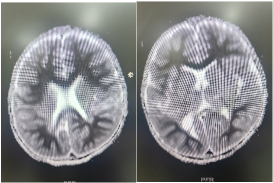
2 year old boy presented to Medanta, the medicity with history of fever, loose stools since 3 days and posturing since last 1 day. Admitted outside, loaded with levetiracetam, phenytoin and valproate and sent to medanta in view of persistent posturing. The child was intubated in view of poor GCS and started on midazolam infusion and later Ketamine infusion was added. Seizures controlled clinically and EEG also confirmed the same. MRI Brain done s/o- symmetrical diffusion restriction involving the bilateral frontoparietal, periventricular and deep white matter with no grey matter involvement s/o ALERD . CSF study was suggestive of- normal biochemistry 2 cells with negative PCR .The child was given IVIG and pulse methyl prednisolone in view of inflammatory pathology. Autoimmune enphalitis work up was negative. The child improved gradually , extubated and discharged.
DISCUSSION
It is a clinical-radiological syndrome and is one of the infection-associated encephalopathy syndromes seen in childhood. It has a low mortality rate but has a higher incidence (>2/3rd) of neurologic sequelae. It has been reported mainly from East Asia. In a series of 44 patients from Nagoya University, Japan, causative pathogens isolated were human herpesvirus-6, adenovirus, rotavirus, influenza virus, Mycoplasma pneumoniae, Enterovirus type Coxsackievirus A6, Escherichia coli O157:H7, and Streptococcus pneumonia.
Fig 1, 2: MR images showing diffusion restriction in Bilateral symmetrical periventricular and deep white matter.

Figure 1
