Mechanical Failure and Complications Following TFNA Fixation in a Comminuted Intertrochanteric Femur Fracture
Mechanical Failure and Complications Following TFNA Fixation in a Comminuted Intertrochanteric Femur Fracture
Dr Saif Saeed Almehrzi*
*Correspondence to: Dr Saif Saeed Almehrzi, Consultant orthopedic, sheikh Shakhbout Medical City (SSMC), Abu Dhabi, United Arab Emirates.
Copyright
© 2025 Dr Saif Saeed Almehrzi is an open access article distributed under the Creative Commons Attribution License, which permits unrestricted use, distribution, and reproduction in any medium, provided the original work is properly cited.
Received: 23 June 2025
Published: 01 July 2025
Mechanical Failure and Complications Following TFNA Fixation in a Comminuted Intertrochanteric Femur Fracture
Patient Information:
Name: X X
Date of Birth: 01/01/1950
Sex: Female
Medical History: Non-contributory
Mechanism of Injury: Mechanical fall at home
Presentation:
The patient, a 75-year-old female, presented to the emergency department following a fall at home. Radiographic evaluation revealed a comminuted intertrochanteric femur fracture on the right side. The patient was admitted for surgical management after appropriate medical and anesthetic clearance.
Surgical Intervention:
The patient underwent internal fixation using a Trochanteric Fixation Nail Advanced (TFNA). Intraoperatively, fracture reduction was suboptimal, with mild varus malalignment. The entry point for the nail was slightly lateral, and the cephalic screw was inserted in an inferior-anterior position. Intraoperative fluoroscopy showed acceptable placement, although the exact position of the screw could not be fully assessed.
Postoperative Imaging and Diagnosis:
Postoperative X-rays were inconclusive due to suboptimal views. To evaluate screw position and fracture alignment more accurately, a CT scan was performed. The CT revealed that the cephalic screw had cut through the femoral neck, protruding medially outside the cortex, and continued into the femoral head—confirming a case of cut-through.
Multidisciplinary Review and Management Plan:
The case was reviewed in a multidisciplinary orthopedic surgery meeting. Given the evidence of mechanical failure, the decision was made to proceed with revision surgery.
Planned Revision:
Anatomical fracture reduction
Use of a medial-posterior nail entry point
Placement of the cephalic screw in a central–central position within the femoral head to optimize mechanical stability
Discussion:
Complications Associated with TFNA Fixation
This case illustrates a classic set of complications associated with suboptimal implant positioning and complex intertrochanteric fractures:
1. Cut-through (Confirmed in This Case):
The screw perforates the medial femoral neck and enters the femoral head.
Strongly associated with inferior-anterior screw positioning, as seen here.
Results in loss of fixation and requires revision.
2. Cut-out:
Migration of the screw superiorly out of the femoral head.
Risk is elevated with eccentric screw placement and poor bone quality.
Leads to painful collapse and loss of fixation.
3. Medial Migration:
Progressive medial movement of the screw into the pelvis.
Occurs with non-central screw positioning and inadequate bone support.
Can result in severe complications including pelvic perforation.
4. Implant Breakage:
Mechanical failure due to repeated stress, particularly at the screw-nail interface.
More likely when fracture is unstable or reduction is inadequate.
Requires implant removal and replacement.
5. Periprosthetic Fracture:
Fractures occurring around or below the tip of the nail.
A lateral entry point may create stress risers, especially in osteoporotic bone.
6. Nonunion:
Failure of the fracture to heal due to instability and poor implant mechanics.
Can lead to chronic pain and disability, necessitating reoperation.
7. Malunion:
Healing in a malaligned position, such as varus or rotational deformity.
Can impair gait and cause limb length discrepancy.
8. Infection:
Although not directly linked to implant positioning, infection remains a general surgical risk.
Can complicate healing, particularly if revision surgery is needed.
9. Avascular Necrosis (AVN):
Disruption of femoral head blood supply, potentially exacerbated by multiple screw placements or medial perforation.
May require conversion to total hip arthroplasty in advanced cases.
Conclusion
This case highlights the critical importance of accurate implant placement in managing comminuted intertrochanteric femur fractures. Inferior-anterior positioning of the cephalic screw and a lateral nail entry point significantly increase the risk of complications such as cut-through, cut-out, and implant failure. Early identification through imaging and prompt revision with improved technique (medial entry, central screw placement) are key to optimizing outcomes and minimizing morbidity.
Post fall xray :
Fig 1
Fig 2
Post first surgery :
Fig 3
Fig 4
Fig 5
Revision surgery :
Fig 6
Fig 7

Figure 1
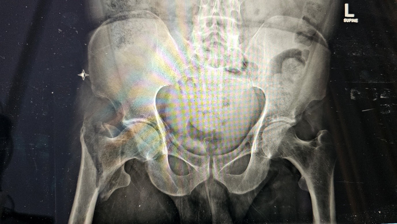
Figure 2

Figure 3
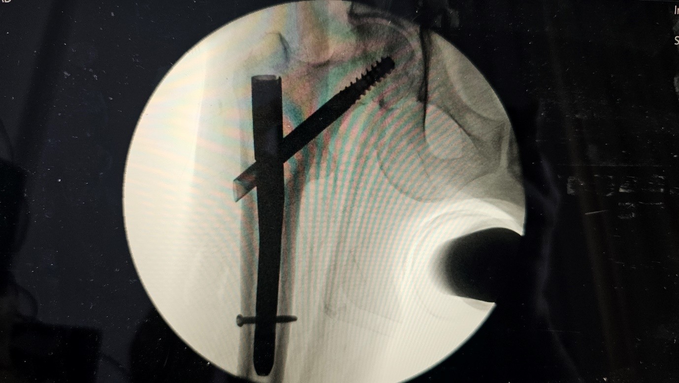
Figure 4

Figure 5

Figure 6

Figure 7