Real-World Experience of Bulkamid® Treatment for Women with Stress Urinary Incontinence
Real-World Experience of Bulkamid® Treatment for Women with Stress Urinary Incontinence
Panayoti Bachkangi 1,2, *, Tajinder Khera 3, Mohamed Saleh 4, Shaima Ibrahim 5,
Maisa Salman 6 Bee K Tan 2
1. United Derby and Burton NHS Foundation Trust.
2. University of Leicester.
3. Hull University Teaching Hospitals NHS Trust.
4. United Lincolnshire Hospitals NHS Trust.
5. Whittington Health NHS Trust.
6. Aman Hospital, Doha, Qatar.
*Correspondence to: Panayoti Bachkangi, United Derby and Burton NHS Foundation Trust.
Copyright
© 2023 Panayoti Bachkangi. This is an open access article distributed under the Creative Commons Attribution License, which permits unrestricted use, distribution, and reproduction in any medium, provided the original work is properly cited.
Received: 16 August 2023
Published: 01 September 2023
Abstract
Introduction and Hypothesis: Polyacrylamide hydrogel (Bulkamid®) urethral injection, is a sur-gical option for the management of stress urinary incontinence (USI). However, very few studies have focused on real-world evidence of Bulkamid® in clinical practice and whether success rate or complications differs from the clinical trial setting. We therefore aimed to review a series of consecutive cases treated with Bulkamid® to investigate outcomes, complications, and to deter-mine predictive factors for treatment success. Methods: A regional, retrospective, observational study, that included all the women who underwent Bulkamid® over a four-year period in 4 op-erating centres (2016-2020). Outcomes were assessed within 12 weeks of the procedure using the British Society of Urogynaecology (BSUG) validated questionnaires. Results: In total, 158 women had Bulkamid®, of which 85% had it for the first time. Almost a third of women (31%) were managed under gynaecology, 32% under urology, and 35% under urogynaecology. The subjec-tively reported success rate was 66% (n=104/159). Of those women with successful outcomes, 80% (n=83/104) had complete success and 20% (n=21/104) moderate success. However, 3.2% of women had a post-operative complication. On univariate analyses, higher success rate was observed in women managed under urogynaecology (74.6%) or gynaecology (71.4%), in procedures per-formed under local anaesthetic, in non-smokers and women aged over 60 years, while BMI did not affect the success rate. There variables were not confirmed as significant predictors on mul-tivariate analyses. However, the year in which the procedure was performed significantly pre-dicted success rate in the multivariate analyses and surgeons-in-training who performed only 1 procedure had a 100% failure rate, demonstrating the importance of supervision prior to sign-off to improve operative outcome. Conclusions: Our real-world evaluation demonstrates significant symptomatic improvement, with minimal complications provided the procedure is performed frequently by specialists.
Keywords: Bulking agents; urinary incontinence; surgical management.
Real-World Experience of Bulkamid® Treatment for Women with Stress Urinary Incontinence
Introduction
Stress urinary incontinence (SUI) is a problem that adversely effects quality of life for millions of women worldwide. Stress incontinence is involuntary leakage from the urethra with effort or physical exertion, or on sneezing or coughing [1],
The most recent National Institute for Health and Care Excellence (NICE) guide-lines for SUI recommend first-line treatment with physiotherapy and pelvic floor training exercises as conservative management [2]. Following this, surgical manage-ment is recommended with mid-urethral slings (mesh repair), autologous fascial slings or urethral bulking agents. However, there have been significant safety concerns about mesh repairs in recent years which has lead to a pause of their use [3]. Consequently, there has been a shift in practice towards bulking agents which are less invasive. A 2017 Cochrane review recommended bulking agents as a treatment option in women for whom general anaesthetic would be too high risk, but advised that definitive surgery should be offered to all low-risk women [4]. Nevertheless, bulking agents remain a popular surgical option among surgeons due to their indication for fragile and surgically high-risk patients [5].
Bulkamid® is a polyacrylamide hydrogel (PAHG), non-toxic, non-resorbable bulking agent, which is recommended for SUI or mixed urinary incontinence (MUI) [6]. Bulking agents are injected into the submucosa of the mid-urethra or neck of the blad-der, to induce urethral coaptation, which increases the urethral mass effect and stimu-lates contractility and stretch of the rhabdosphincter muscle fibres, to counteract SUI [7]. Bulking agents are injected peri-urethrally (parallel to) or trans-urethrally (along-side) a cystoscope under local or general anaesthetic [8].
Several systematic reviews and randomised controlled trials have demonstrated efficacy and safety of Bulkamid® treatment, with reduced frequency and severity of complications, compared to mesh repair [4, 9, 10]. Notably, in all these studies, the surgeons had completed their training and were experienced in performing the pro-cedure [10].
The real-world evidence of Bulkamid® in clinical practice is limited and is unclear whether the success rate or complications differs from the clinical trial setting. We therefore aimed to review a series of consecutive cases treated with Bulkamid® to in-vestigate outcomes, complications, and to determine predictive factors for treatment success.
Methods
We conducted a regional, retrospective, observational study, which included a convenience sample of consecutive women who underwent a Bulkamid® procedure performed for stress or mixed urinary incontinence. The study ran from the introduction of Bulkamid treatment to the trusts and continued over a four year period, from January 2016 to December 2019 inclusive. There were 4 operating centres: Pilgrim Hospital (Boston), Lincoln County Hospital (Lincoln), Diana Princess of Wales (DPOW) and Scunthorpe General Hospital (SGH)) and 13 operating surgeons. All surgeons (n=13) received the same training for performing the Bulkamid procedures, which comprised competency-based training under supervision with the surgeon than signed off for in-dependent practice.
Incontinence investigations
All women were seen in the urinary incontinence clinic by a urology specialist (urologist or urogynaecologist) and had a full history, and clinical examination. The urodynamics investigations were performed according to NICE guidelines [2], and were used to confirm the diagnosis of mixed or stress urinary incontinence. Women were provided with appropriate treatment options including conservative, medical and sur-gical management, for which the latter included mid-urethral slings and/or Bulkamid® treatment. Moreover, as per local and national guidelines, the plans of management should be discussed in the pelvic multidisciplinary team meeting, including urology and/or urogynaecology consultants, urology nurse specialist and a pelvic floor specialist physiotherapist. The MDT may also include a geriatrician, occupational therapist and colorectal surgeons [2].
Operative procedure:
Written informed consent was obtained from all women prior to the Bulkamid® procedure. The procedure was performed under local or general anaesthetic dependent on patient and operating surgeon factors. All women were treated with antibiotic prophylaxis immediately prior to the procedure. The procedure was performed in the lithotomy position and a cysto-urethroscope was used. Bulkamid® treatment was in-jected through the specified rotatable sheath, in accordance with manufacturers guide-lines. 2 mls of Bulkamid® was injected under direct visualisation in the 2, 5, 7 and 11 ‘o’ clock positions of the mid-urethral mucosa. Standard operating procedures and ap-propriate training from a Bulkamid® representative minimised intra-operative varia-tion. Following the procedure women were discharged on the same day after having a successful attempt of micturition.
Outcomes:
Women were followed-up in clinic within 12 weeks Bulkamid® treatment. The success of the Bulkamid® treatment was determined using the British Society of Urogynaecology (BSUG) Patients Global Impression of Improvement for Incontinence (PGII) [11], a validated questionnaire and standardised measure of procedural success for any surgical management for USI. Patients completed the urinary continence ques-tionnaire within 12 weeks of the procedure. Complete success was defined as ‘very much better’ symptomatic improvement in urinary incontinence on the questionnaire; moderate success was defined as ‘much better’ or ‘a little better’; and unsuccessful was defined as ‘no change’ or symptoms which were ‘a little worse’, ‘much worse’, or ‘very much worse’.
For the purposes of this paper, two outcome systems were used, the first com-paring ‘any improvement’ (complete or moderate combined) to ‘unsuccessful’ proce-dures (two possible outcomes). The second outcome system was comparing ‘completely successful’, ‘moderately successful’ and ‘unsuccessful’ procedures (three possible outcomes).
Data was collected directly from the BSUG national dataset.
Safety outcomes were frequency of adverse events and immediate, or late surgical complications within 12 weeks of the procedure. Adverse events and complications were collected from the patient notes and clinic letters.
Determinants of success:
A number of patient, pre-operative and operative factors were assessed. Patient factors included age (years), BMI (kg/m2), previous Bulkamid® treatment (primary or repeat procedure) and smoking status (yes or no). Pre-operative factors included physiotherapy (yes or no), patient information leaflet (yes or no), case discussion in a multidisciplinary team meeting (yes or no) and which specialty the women was man-aged under (gynaecology, urology or urogynaecology). Operative factors included an-aesthetic used (local or general), surgical time, centre procedure was performed at (Lincoln, Boston, DPOW, or SGH) and year procedure was performed (2016-2020).
Statistical analysis
Descriptive statistics were provided for baseline characteristics. The distribution of data was found to be non-parametric on gaussian distribution. The chi square test and z tests were used to determine statistical significance between nominal groups. The Mann Whitney U test was used to compare continuous variables with nominal outcome groups. Multinomial logistic regression analyses were performed in SPSS to identify independent predictors of outcome success. All statistical analyses were performed in SPSS v22 and graphs were created in GraphPad Prism. Statistical significance was set at p<0.05.
Ethics:
Data used in this observational study was collected as part of the national Bulka-mid® audit. All data was anonymised at the point of data collection.
Results
Baseline characteristics
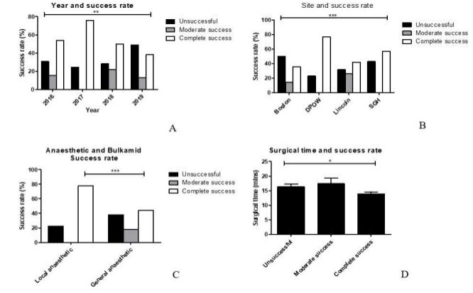
A total of 158 women had a Bulkamid® procedure performed between January 2016 and January 2020 across four sites in the Lincolnshire region (Boston n=42/158, DPOW n=52/158, Lincoln n=57/158, and SGH (n=7/158).
The majority of Bulkamid® procedures in this cohort were performed by a con-sultant (97%). Of the 13 operating surgeons, 4 performed a high number of procedures (range 21 – 41 procedures), 3 had performed a moderate number of procedures (range 7-11 procedures) and 6 had performed only 1 procedure. Surgeons who performed a high number of procedures (n=125 procedures) had a 54% complete success rate, 15% moderate success rate and a 31% failure rate. Surgeons who performed a moderate number of procedures (n=27 procedures) had a 63% complete success rate, 7% moderate success rate and a 30% failure rate. Surgeons who only performed 1 procedure (n=5 procedures) had a 100% failure rate and all of these cases were performed by a surgical registrar.
Median patient age was 55 years (46-69 years IQR), median BMI was 30 (26-34 IQR) and 6% (n=9/158) were smokers (56%, n=88/158 were non-smokers and smoking status was unknown for 39% (n=61/158).
For 85% (n=135/158) of women, this was their first Bulkamid® procedure, whereas 14% (n=11/158) had undergone one or more previous Bulkamid® procedure(s). In total, thirty one per cent (n=49/158) of women had undergone a previous urological procedure for urinary incontinence, including Bulkamid® (n=11), AP repair (n=1), Birch colpo-suspension (n=2), botox (n=3) or mesh (n=1).
Pre-operative Management
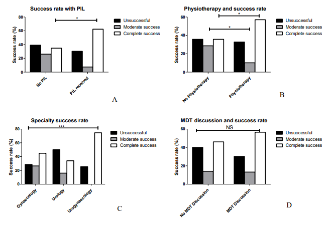
Thirty one per cent (n=49/158) of women were managed under gynaecology, 32% (n=50/158) under urology, and 35% (n=59/158) had joint care under urogynaecology.
Pre-operatively, 81% (n=128/158) of women received physiotherapy, 67% (n=106/158) had their case discussed in a multidisciplinary team and of these, 100% were managed as per the multi-disciplinary team (MDT) plan, and 67% (n=106/158) received a patient information leaflet about Bulkamid® treatment prior to the procedure.
Patient factors:
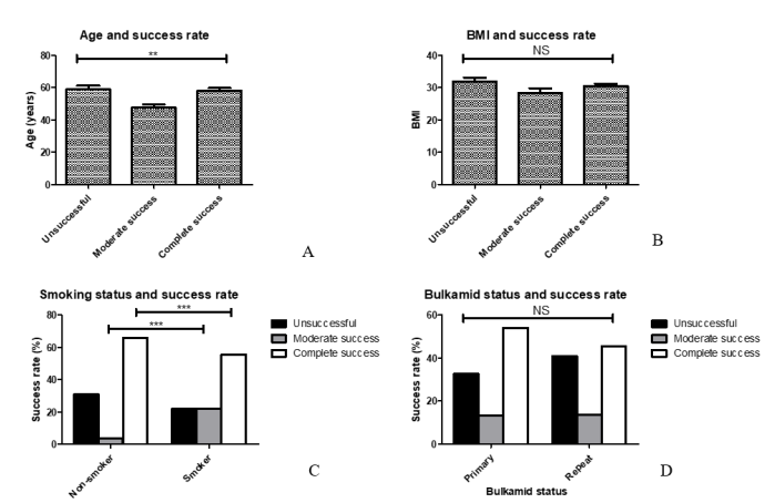
Non-smokers were significantly more likely to attain a completely successful outcome compared to smokers (65.9% vs 55.6% respectively, p<0.0001) (Figure 1c.).
Age was significantly (p=0.029) higher in completely successful procedures (me-dian age = 57 years) compared to moderately successful procedures (median age = 45 years). However, there was no significant difference in age of woman between the completely successful procedures and those which were unsuccessful (median = 57 years) (Figure 1a). BMI did not differ between women with successful and unsuccessful outcomes (Figure 1b.) and whether this was their first Bulkamid® or a repeat did not affect the success rate (Figure 1d.).
Figure 1. Patient factors for the success of Bulkamid®. a. Advanced age (median= 57 years) was associated with complete success in comparison to younger patients (p=0.029). b. The BMI of the patients does not seem to be a predictive factor for the success of the operation (p >0.05). c. Pa-tients who did not smoke had significantly successful outcome (p=0.0001) in comparison to smokers. d. Bulkamid success is not influenced by whether the procedure is primary or repeat (p >0.05).
Surgical outcome and complications
Median follow-up time was 6 weeks (6-12 weeks IQR). The Bulkamid® procedure was unsuccessful in 34% (n=54/158) of cases and was successful in 66% (n=104/159) of cases. Of those women who had a successful outcome, this was defined as a complete success in 80% (n=83/104) of cases and a moderate success in 20% (n=21/104) of cases. Surgeons who only performed one Bulkamid® procedure had a 100% failure rate. 3.2% of women had a post-operative complication at follow-up (n=2 worsening urgency, n=1 urinary retention, n=1 new urgency, n=1 dysuria and n=1 intermittent haematuria).
Factors determining treatment success
Adopting the two-tier outcome system (successful vs unsuccessful), Bulkamid® success rate was significantly higher in women managed under urogynaecology (74.6%) or gynaecology (71.4%) compared to women managed under urology (50.0%) (p=0.016) (Figure 2c.). Also, in centres where more procedures had been performed, there was a trend for a significantly higher success rate (p=0.048) (Figure 3b.).
Success rate was significantly higher in 2017, when n=37 procedures were per-formed, compared to any other year (75.7%, p=0.009), although the highest number of procedures were performed in 2018 (n=60 procedures) and 2019 (n=47 procedures) (Figure 3a.).
The sites that performed the highest number of procedures (n= 52 at DPOW and n=57 at Lincoln) had significantly higher complete (76.9%) and moderate success rates (26.3%) respectively (p<0.0001), compared to Boston (n=42 procedures) and SGH (n=7 procedures) (Figure 3b.).
A completely successful outcome was more likely to be attained in women man-aged under joint care by urogynaecology (74.6% success rate, p<0.0001) compared to women under urology (34.0% success rate) or gynaecology (44.9% success rate) alone (Figure 2c.). Women managed under gynaecology were more likely to attain a moder-ately successful outcome (26.5%, p<0.0001) compared to those managed under urology (16%) or joint care (0%).
Figure 2. Pre-operative management and Bulkamid® success. a. Offering PIL preoperatively is a significant factor for a successful outcome (p=0.0001). b. Physiotherapy is the first line of man-aging SUI and is associated with significantly successful surgical outcomes (p=0.0002). c. The procedures performed by a specialist in urogynaecology were associate with the highest success rates, followed by those performed by general gynaecologists and urologists (p<0.0001). d. MDT discussions did not influence the surgical outcomes (p.0.05) but they still remain an essential preoperative step.
Operative procedure
Local anaesthetic (with or without sedation) was used in 25% (n=40/158) of cases, and general anaesthetic was used in 75% (n=118/158) of cases. On average, surgical time was 13 minutes (11-18 minutes IQR). Shorter surgical time was associated with com-plete success in comparison to longer times (13 minutes vs 19 minutes; p=0.0479) (Figure 3d.).
Figure 3. Operative success factors: a. The procedures performed in 2017 were the most effica-cious (p=0.009) with complete satisfaction among successful patients. This could possibly be due to the enhanced training and supervision the surgeons have been offered. b. Busier units, like DPOW and Lincoln, where many procedures are performed, are a significant predictor of the success of Bulkamid® (p<0.0001). This suggests that the higher the exposure to a surgical proce-dure, the more likely the results to be positive. c. Procedures performed with local anaesthesia were associate with higher success rates (p<0.0001), possibly because the surgeons performing them are very experienced. d. Shorter surgical times were a positive predictor of the surgical outcome (p=0.0479). Again, this could be due to the fact that more experienced surgeons can perform the procedures relatively faster.
Pre-operative factors
Pre-operative physiotherapy made attainment of a completely successful outcome significantly more likely compared to no pre-operative physiotherapy (57.0% vs 35.7%, p=0.020), as did receiving an information leaflet beforehand (62.3% vs 34.8%, p=0.001) (Figure 2a. and 2b.).
Peri-operative factors
Interestingly, a completely successful outcome was more likely to be attained with local anaesthetic (77.5% complete success rate) compared to general anaesthetic (44.1% complete success rate, p<0.0001) (Figure 3c.). General anaesthetic also had a significantly higher failure rate (38.1% failure rate) compared to local anaesthetic (22.5%, p<0.0001).
Logistic regression
Multinomial logistic regression was performed for the three tier outcome system and analyses showed that only year in which procedure was performed was a signifi-cant predictor; the likelihood of achieving a moderate outcome significantly increased from year 1 (2016) to year 3 (2018) of procedures (Figure 3a.).
Multinomial logistic regression was also performed for the two tier outcome sys-tem and showed that none of the assessed factors predicted likelihood of a successful or unsuccessful outcome.
Discussion
Our real-world, regional evaluation of the Bulkamid® therapy demonstrates sig-nificant symptomatic improvement in urinary incontinence, with minimal complica-tions from 3 months of follow-up in our cohort. Our improvement rate of 66% is lower than that reported by Pai et al. of 82.8% which persisted over time, but pre-operative severity of urinary incontinence is an important predictor of outcome [12]. For example, Giammo et al. found a lower success rate with Bulkamid® treatment of 36.1% and a higher complication rate with 8.2% suf-fering post-operative urinary retention, but this was in a cohort with severe urinary incontinence (p=0.008) [13]. They showed that more severe urinary incontinence was significantly associated with a poorer outcome (p=0.008). Other studies have shown mixed results with improvement rates of 67-88% at 24 months follow-up, further cor-roborating patient factors in determining outcome [13, 16]. A recent systematic anal-ysis, though, has suggested short-term improvement rates of 30-90% with Bulkamid® and 42%–70% on the long term [16], concluding that our findings are very satisfactory.
The only significant predictor on multivariate analysis was year in which proce-dure was performed with a higher success rate in 2018 compared to 2016, suggesting that increased frequency of performing the Bulkamid® procedures improves the pa-tient-reported outcome. Further, surgeons who performed only 1 procedure in this cohort had a 100% failure rate whereas surgeons who performed a moderate or high number of procedures had a 30-31% failure rate. This suggests the outcome with Bulkamid® treatment improves with increased frequency of procedures performed. Surgeons new to the Bulkamid® procedure likely require observation for more than one procedure prior to sign-off. This further is substantiated by the low frequency group being comprised solely of surgical registrars. Surgeons in training need to be supervised and supported with Bulkamid procedures during their training period, to ensure com-petency is achieved, particularly as the number of Bulkamid® procedures undertaken will increase in their consultant years.
Although multivariate analyses did not confirm the significant predictors identi-fied from the univariate analyses in our study, the small convenience sample used likely accounts for this. In the discussion below, we have outlined the univariate predictors in relation to scientific literature, as these findings warrant further exploration in a larger cohort.
In our series, age was lower in cases where a moderately successful outcome was achieved but was equivalent in completely successful and unsuccessful procedures. Evidence shows that women aged over 60 years have a higher likelihood of successful outcome, attaining over 90% success rate [15[. This highlights the importance of opti-mising conservative and medical management approaches and offering Bulkamid® treatment in older women for whom invasive procedures may be too high risk [17]. Further, smoking reduced likelihood of a successful outcome, hence patients should be counselled about this prior to undertaking the Bulkamid® procedure. Importantly, in our study, previous Bulkamid® treatment did not affect the outcome [8, 10].
We also identified pre-operative and operative factors which affected likelihood of success. Patients managed under urogynaecologists were more likely to have a com-pletely successful outcome demonstrating, the importance of MDT input and man-agement with Bulkamid® treatments. Similar superiority in the clinical outcomes has been demonstrated before in gynaecologic oncology, where better results are reached when patients are managed by subspecialised teams [18-20].
Also, women who received a patient information leaflet and physiotherapy pre-operatively were more likely to achieve to a completely successful outcome as op-posed to a moderately successful outcome. This demonstrates the need to adhere to NICE guidelines with respect to optimising conservative management prior to opera-tive management, and managing expectation and patient understanding through pa-tient information leaflets [2].
Cases performed with local anaesthetic were more likely to be completely suc-cessful than those with general anaesthetic. Reasons for this include disparate patient risk profiles, which would be higher in the general anaesthetic group, but also proce-dural competence, as practitioners who performed more procedures transitioned from general to local anaesthetic over time, and a higher success rate would be expected to follow with this. This is also suggested by our regression analyses showing an increase in moderately successful procedures over time, which reflects introduction of more surgeons into the operative pool, and suggests that frequency of performing Bulkamid® treatments, may predict complete vs moderately successful outcomes. Finally, com-pletely successful procedures were also shorter compared to moderately successful or unsuccessful procedures, which further supports the importance of operative frequency and training, to optimise outcomes, and highlights an area for further investigation.
Notably NICE does not recommend “bulking agents” unless all the alternative recommended surgical options, including the retropubic mid-urethral mesh slings, are not suitable or acceptable [2]. It is understandable that bulking agents, in general, have been avoided in the past due to insufficient evidence of their efficacy and durability. However, there are different types of bulking agents, like autologous fat, carbon beads, porcine dermal implant, silicon particles calcium hydoxylapate, and collagen [4]. Un-derstandably, it is not expected that all these agents would offer similar results, while generalising the lack of suitability to all “bulking agents” does not do fairness to PAHG, especially as the preliminary evidence previously has shown promising results with regards its success [10]. Bulking agents have been recommended by the International Consultation on Incontinence committee among the first-line of surgical options [21].
In recent studies comparing TVT to Bulkamid®, the tape showed indeed some superiority in patient satisfaction regarding their urinary symptoms [9, 22]. Yet, the rate of complications and associated pain was considerably lower with Bulkamid®, sug-gesting that PAHG is an efficacious alternative option of management especially in pa-tients with multiple comorbidities, where minimally invasive procedures are preferred to reduce surgical risk. Hence, Bulkamid® has shown advantageous outcomes and effect and safety in comparison to other bulking agents [16].
This study offers a real-world assessment of patients undergoing Bulkamid® procedure across four centres, which to our knowledge, has not previously been re-ported on. Limitations of this study include the retrospective data collection, the lack of longer-term follow-up data and lack of information about severity of incontinence prior to performing the Bulkamid® procedure and comorbidity information. Also, the sur-gical outcomes were not measured at a fixed time point (within 12 weeks post-operatively) rendering it difficult to compare the effect of time after surgery on determination of the outcome observed. However, the timing of the follow up at 12 weeks is based on the BSUG recommendations. As for the use of Bulkamid®, this is a bulking agent commonly used in the U.K., and the authors did not have the choice between that agent and any other. Finally, success was measured according to patient satisfaction which is an important albeit subjective measure; ideally, the success of the procedure should have been measured both subjectively, by questionnaires and objec-tively by urodynamics, and the follow up period should have been extended [23]. However, objective measures of improvement were not routinely adopted by specialists across the four trusts and the BSUG’s recommendation for assessment of pa-tient-reported outcome measures was adhered to in this study. Finally, the study was not powered to demonstrate effectiveness or safety profiles for Bulkamid treatment and there was no control group available for comparison purposes. As multivariate analyses did not confirm the univariate factors, evaluation in a larger cohort form additional centres is warranted to explore predictors of success with Bulkamid® treatment. Con-sequently, we recommend a prospective randomized control trial to reassess the treatment outcome for this modality in the future. Nevertheless, this study aimed to evaluate the re-al-world success of the procedure with a focus on subjective outcomes as an important measure of this.
Conclusion
PAHG (Bulkamid®) is an effective bulking agent for the surgical management of USI. Our real-world assessment demonstrates comparable success rates to clinical trial data, but supports that surgeons-in-training require observation and need to perform the procedure frequently to attain good surgical outcomes. Long-term follow up as-sessments, including objective evaluations, are recommended for future studies to evaluate the long-term success of the procedure.
References
1. Haylen, B.T., et al., An International Urogynecological Association (IUGA)/International Continence Society (ICS) joint report on the terminology for female pelvic floor dysfunction. Neurourol Urodyn, 2010. 29(1): p. 4-20.
2. NICE National Institute for Health and Care Excellence: Urinary incontinence and pelvic organ prolapse in women: man-agement. 2019. 76.
3. Willet K., M.K., Recommendation of the Mesh Pause Clinical Advisory Group to Medical Directors and Surgical Teams, N.I.a.N. England, Editor. 2018. p. 10.
4. Kirchin, V., et al., Urethral injection therapy for urinary incontinence in women. Cochrane Database Syst Rev, 2017. 7: p. CD003881.
5. Serati, M., V. Mancini, and M. Balzarro, Urethral bulking agents for the treatment of female stress urinary incontinence. Int Uro-gynecol J, 2020. 31(8): p. 1493-1494.
6. Brosche, T., et al., Seven-year efficacy and safety outcomes of Bulkamid for the treatment of stress urinary incontinence. Neurourol Urodyn, 2021. 40(1): p. 502-508.
7. Klarskov, N. and G. Lose, Urethral injection therapy: what is the mechanism of action? Neurourol Urodyn, 2008. 27(8): p. 789-92.
8. Hussain, S.M. and R. Bray, Urethral bulking agents for female stress urinary incontinence. Neurourol Urodyn, 2019. 38(3): p. 887-892.
9. Itkonen Freitas, A.M., et al., Quality of life and sexual function after TVT surgery versus Bulkamid injection for primary stress urinary incontinence: 1 year results from a randomized clinical trial. Int Urogynecol J, 2021. 32(3): p. 595-601.
10. Kasi, A.D., et al., Polyacrylamide hydrogel (Bulkamid(R)) for stress urinary incontinence in women: a systematic review of the literature. Int Urogynecol J, 2016. 27(3): p. 367-75.
11. Srikrishna, S., D. Robinson, and L. Cardozo, Validation of the Patient Global Impression of Improvement (PGI-I) for uro-genital prolapse. Int Urogynecol J, 2010. 21(5): p. 523-8.
12. Pai, A. and W. Al-Singary, Durability, safety and efficacy of polyacrylamide hydrogel (Bulkamid((R))) in the management of stress and mixed urinary incontinence: three year follow up outcomes. Cent European J Urol, 2015. 68(4): p. 428-33.
13. Giammo, A., et al., Urethral bulking with Bulkamid: An analysis of efficacy, safety profile, and predictors of functional outcomes in a single-center cohort. Neurourol Urodyn, 2020. 39(5): p. 1523-1528.
14. Ghoniem, G., et al., Cross-linked polydimethylsiloxane injection for female stress urinary incontinence: results of a multi-center, randomized, controlled, single-blind study. J Urol, 2009. 181(1): p. 204-10.
15. Toozs-Hobson, P., et al., Two-year follow-up of an open-label multicenter study of polyacrylamide hydrogel (Bulkamid(R)) for female stress and stress-predominant mixed incontinence. Int Urogynecol J, 2012. 23(10): p. 1373-8.
16. Hoe, V., et al., Urethral bulking agents for the treatment of stress urinary incontinence in women: A systematic review. Neurourol Urodyn, 2021.
17. Vecchioli-Scaldazza, C.V., et al., Polyacrylamide hydrogel (bulkamid(R)) in female patients of 80 or more years with urinary incontinence. Int Braz J Urol, 2014. 40(1): p. 37-43.
18. du Bois, A., et al., Variations in institutional infrastructure, physician specialization and experience, and outcome in ovarian cancer: a systematic review. Gynecol Oncol, 2009. 112(2): p. 422-36.
19. Fung-Kee-Fung, M., et al., The optimal organization of gynecologic oncology services: a systematic review. Curr Oncol, 2015. 22(4): p. e282-93.
20. Vernooij, F., et al., The outcomes of ovarian cancer treatment are better when provided by gynecologic oncologists and in specialized hospitals: a systematic review. Gynecol Oncol, 2007. 105(3): p. 801-12.
21. Recommendation of the International Scientific Committee: surgery for urinary incontinence in women., in 6th International Consultation on Incontinence, C.L. Abrams P, Wagg A, Wein A Editor. 2017: Paris, France. p. 2570–1.
22. Itkonen Freitas, A.M., et al., Tension-Free Vaginal Tape Surgery versus Polyacrylamide Hydrogel Injection for Primary Stress Urinary Incontinence: A Randomized Clinical Trial. J Urol, 2020. 203(2): p. 372-378.
23. Lo, T.S., et al., Urodynamics mixed type urinary incontinence with advanced pelvic organ prolapse, management and out-comes. Sci Rep, 2020. 10(1): p. 1944