Methemoglobinemia: A Case Diagnosed in the Early Days of Life
Methemoglobinemia: A Case Diagnosed in the Early Days of Life
Dr Latifa Mouttawa, M.D., D.CH, M.R.C.P.CH 1, Dr. Shiva, M.B.Ch.B., M.R.C.P. Ch 2,
Dr. Ban F. Refaat, M.B.Ch.B.,C.A.B.S. Ph.D 3
1. Consultant in Paediatrics, Paediatrics Department, Sohar hospital, Sohar, Sultanate of Oman.
2. Senior Specialist in Paediatrics, Paediatrics Department, Sohar hospital, Sohar, Sultanate of Oman.
3. Specialist in Paediatrics, Paediatrics Department, Sohar hospital, Sohar, Sultanate of Oman.
*Correspondence to: Dr Latifa Mouttawa, M.D., D.CH, M.R.C.P.CH. Consultant in Paediatrics, Paediatrics Department, Sohar hospital, Sohar, Sultanate of Oman.
Copyright
© 2023 Dr Latifa Mouttawa. This is an open access article distributed under the Creative Commons Attribution License, which permits unrestricted use, distribution, and reproduction in any medium, provided the original work is properly cited.
Received: 23 September 2023
Published: 05 October 2023
Methemoglobinemia: A Case Diagnosed in the Early Days of Life
Introduction
Methemoglobin (MetHb) is altered state of hemoglobin (Hb) in which the ferrous (Fe2+) irons of heme are oxidized to the ferric (Fe3+) state.
The ferric hemes of MetHb are unable E to bind oxygen (O2).
Thus, oxygen dissociation curve is left-shifted, making it more difficult to release O2.
Methemoglobinemia either a congenital type which is due to diminished enzymatic reduction of MetHb back to functional Hb, affected patients appear cyanotic but are generally asymptomatic or acquired type which is due to specific drugs that cause oxidation of Hb to MetHb more rapidly than MetHb is reduced to Hb. which can be fatal. (2, 3, 7, 20)
Here we report a case of congenital Methemoglobinemia in the early days of life, which is a very uncommon.
We discuss the clinical picture, way of diagnosis and review of literatures.
Key words: Methemoglobin (MetHb).
The Case
This patient is a newly born baby female 40 + weeks –spontaneous vaginal delivery of 28years old mother G2P1, defaulter butno maternal illness.
Apgar score at 1 and 5 mints was 8,9respectively.
Anomaly scan- normal. High vaginal swab culture and sensitivity was negative.
Then after about 5 hours of birth the baby developed tachypnea with cyanosis, so he was shifted to SCBU unit for further management.
Saturation was 87 in room air and reached 96% on 3 liters nasal pronge O2.
She has also mild nasal flaring, mild dyspnea, cyanosis +, no jaundice.
Chest – good air entry in both sided. HeartS1, S2normal, noadded sounds.
Abdomen soft, no organomegaly, normal external genitalia.
Anterior fontanelle at the level, Moro reflex –normal.
Red reflex +ve. Hip click –normal
Birth length =52 cm, birth weight =3.180 gm, head circumference = 34cm
No congenital anomaly
ABG: as in the table 1: which showed high Met Hb =about 71%
The chest X-ray was normal.
Lab. Investigation:
There was a polycythemia improved with intravenous fluid
Hb=21.10 gm /dl improved to 17.70 ,,,,,,,HCT =69% improve to 57.80%
Reticulocyte count =5.25%
Renal and liver function tests within normal
Neonatal Cord Thyroid stimulating hormone (TSH):=5, 91.
Table 1
The patient was tachypnea, and cyanosed his vital signs as such
Table 2
All possible investigation done including repeated blood gases which showed in the tables attached
Blood gas panel *POC as such
Table 3
Work up sent and case discussed with haematologist in the national center and advice for more investigation so another investigations done as follows:
NGC Genetic testing for haemoglobinopathies (Opinion in Conclusion):
The patient is a carrier of a mild alpha thalassemia, (-a/aa)
GAP PCR for alpha globin cluster indicated that the patient is heterozygous for the (-alpha 3.7 kb) deletion consistent with alpha thalassemiatrait and normal for the (-alpha 4.2,-alpha 20.5,--MED,--SEA) alpha thalassemia deletion.
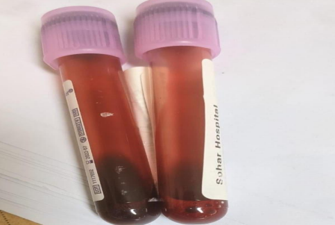
This is the color of the blood sample (Chocolate color)
Figure 1
Peripheral smear showed reaction like infection so medication as follows given:
|
S N |
Medicine |
Strength ,course and frequency |
|
1 |
Benzyl Penicillin inj. 1000000 IuF/1 vial |
310000 IU—BID—5 Days |
|
2 |
Gentamycin Sulphate injection 20Mg/2Ml |
13 Mg ----OD---5 Days |
The patient was kept on Nasal cannula O2 as 3 liters/minute,
NPO on IV fluids, TFR = 70 ml/kg/day, Pass meconium.PT kept initially on Nasal cannula then weaned to room air .Starts feeds next day, suckling well
Progress: the child become active,anterior fontanelle at the level, no temp instability.
He was discharged on the 4th day in stable condition
Advice for follow-up or consult on need to the paediatric emergency unit
Lab on discharge as:
|
Hb |
19 |
|
Hct |
60 |
|
Plt |
288 |
|
Ca |
2,3 |
|
Po4 |
1.9 |
|
ALP |
148 |
|
Albumin |
39 |
|
Na |
140 |
|
K |
4.3 |
|
Cr |
86 |
|
CRP |
12 |
Table 5
Discussion
Methemoglobin (MetHb) is an oxidized derivative of hemoglobin (Hb) in which hem irons is in the ferric (Fe3+) or oxidized state rather than ferrous (Fe2+) or reduced state .(7)
Because methemoglobin is unable to bind (or release )oxygen, the presence of significant amounts of metHb adversely affects oxygen transport .small amounts of methemoglobin normally are formed daily ,associated with the release of oxygen from hemoglobin (auto-oxidation) (7,10,15)
The methemoglobin that is formed rapidly is reduced through the action of RBC NADH methemoglobin reductase (also known as cytochrome b5reductase) so that in normal persons, levels of metHb seldom exceed 1%. A second methemoglobin reductase depends on NADPH as cofactor also is present in RBCs .This enzyme has little function under normal physiologic conditions, but it is greatly activated by the presence of certain redox compounds such as methylene blue, forming the basis for the clinical treatment of methemoglobinemia .
Newborns areparticularly susceptible because fetal hemoglobin is more readily oxidized tothe ferric state than is hemoglobin A andbecause RBC NADH-methemoglobin reductase activity is low during the firstfew months of life (7)
Methemoglobinemia types (1, 7, 8, 9, 11, and 14)
Congenital – diminished enzymatic reduction of MetHb back to functional Hb.
Affected patients appear cyanotic but are generally asymptomatic.
Inherited MetHb: MetHb can be passed down if one or both parents carry a faulty gene that causes problems with the enzyme cytochrome b5 reductase.
Type 1 inherited MetHB: This is also called erythrocyte reductase deficiency and occurs when the red blood cells do not have cytochrome b5 reductase.
Type 2 inherited MetHB: is also called generalized reductase deficiency, and this occurs when many cells in the body do not have the enzyme.
Hemoglobin M disease: This occurs when the hemoglobin protein itself is defective.
Acquired – from specific drugs that cause oxidation of Hb to MetHb more rapidly than MetHb is reduced to Hb. Can be fatal.
Drugs causing MetHb (15, 17, 18): local anesthetics, Benzocaine, nitroglycerine, sodium nitroprusside, phenytoin, sulfonamides, metoclopramide.
When > 10 % (or 1.5 g/dL absolute concentration of MetHb) – central cyanosis occurs.
Symptoms: Cyanosis — a slate-blue color of the skin and mucous membranes, a finding that is due to the different absorbance spectrum of MetHb compared with oxyHb.
MetHb > 20%: respiratory depression, altered consciousness, shock, seizures, and death may occur.
MetHb > 40 % – life threatening.
Congenital methemoglobinemia is due to inherited disorders of hemoglobin structures or to a sever deficiency of NADH methemoglobin reductase activity (4)
The inherited abnormalities of hemoglobin structure that give rise to methemoglobinemia known collectively as the hemoglobin M disorders,are rare autosomal-dominant defects caused by point mutation that alter a single amino acid in the stricter of normal globin (7,12,13)
Only the alpha and gamma globin chain mutations are associated with neonatal methemoglobinemia because these are the goblins that form hemoglobin F. (7,16,20)
Hemoglobin M heterozygotes inheriting alpha or beta globin mutations have lifelong cyanosis, but they are usually asymptomatic. No therapy is needed (and none is possible).
The homozygous state is incompatible with life, NADH –methemoglobin reductase deficiency is a rare autosomal recessive disorder.
Heterozygotes are asymptomatic and do not have methemoglobinemia under normal circumstances .However, if challenged by drugs or chemicals that cause methemogloninemia ,heterozygotes deficits patients may become cyanotic and symptomatic at doses that have no effect in normal persons . (1, 5,6)
Homozygotes have lifelong methemogloninemia levels of 15% to 40% and are cyanotic but otherwise asymptomatic unless exposed to toxic agents.
Normal concentration of MetHb – < 1% of total Hb (auto oxidation of Hb to MetHb occurs spontaneously at a slow rate, each day converting 0.5 to 3 % of the available Hb to MetHb.
This autoxidation, combined with the subsequent reduction of MetHb acts to maintain a steady-state level of MetHb).
Diagnosis of NADH-methemoglobin reductase deficiency is by assay of RBC enzyme activity, aprocedure available only in specialized hematology laboratories.
Cyanosis is first clinically evident when methemoglobin levels reach approximately 10% (1.5 g/dL),but symptoms attributed to hypoxemia and diminished oxygen transport do not appear until levels increase to 30% to 40% of total hemoglobin .Methemogloniemia is not associated with anemia, hemolysis or other hematological abnormalities. (7)
Death occurs at level of 70% orgreater.
Treatment with intravenous methylene blue (1 mg/kg as 1%solution in normal saline) is indicated when methemoglobin levels are greater than 15% to 20 %. (7)
A poor response to methylene blue also is seen in G6PD –deficient persons because this disorder is characterized by suboptimal generation of NADPH. (19)
In general, most infants with hereditary methemoglobinemia are asymptomatic and require no therapy. (7)
Clues for methemoglobinemia:
- -> Cyanosis and low SpO2 in the presence of a normal arterial PO2 by ABG
- -> Presence of “chocolate, dark-red, brownish to blue “colored arterial blood (color does not change with addition of O2) and brown urine
Co-oximeter. On a blood gas, normal PaO2 concentrations are usually found on analysis.
Clinical cyanosis in the presence of normal arterial oxygen tensions is highly suggestive of methemoglobinemia.
Pulse oximetry is inaccurate and unreliable in patients with high methemoglobin fractions. However, an abnormal value in an asymptomatic patient may suggest the presence of an elevated methemoglobin fraction.
The treatment of methemoglobinemia will be (4,6,13,14):
Methylene blue is the first line,it accelerates the enzymatic reduction of methemoglobin by NADPH-methemoglobin reductase and also reduces to leucomethylene blue that, in turn, reduces methemoglobin. This is contraindicated in patients with G6PD deficiency (can cause hemolysis).
Hyperbaric O2 and exchange transfusions can also be utilized.
An asymptomatic patient with a MetHb level <20 % – no therapy other than discontinuation of the offending agent.
Symptomatic patient (or MetHb level is >20 %) – methylene blue (MB). MB – 1 to 2 mg/kg IV over 5 min (total dose should not exceed 7-8 mg/kg – MB can cause dyspnea, chest pain, hemolysis). MB provides an artificial electron transporter for the reduction of MetHb via the NADPH-dependent pathway. The response – rapid; the dose may be repeated in one hour if the level of MetHb is still high 1 hr after the initial infusion. Rebound methemoglobinemia may occur up to 18 hours after MB administration, due to prolonged absorption of lipophilic agents (benzocaine) from adipose tissue. It is reasonable to perform serial measurements of MetHb levels following treatment with MB. MB should not be administered to patients with glucose 6-phosphate dehydrogenase (G6PD) deficiency, since the reduction of MetHb by MB is dependent upon NADPH generated by G6PD (hemolysis). An alternative treatment for these patients – ascorbic acid (2mg/kg). Blood transfusion or exchange transfusion may be helpful in patients who are in shock. Hyperbaric oxygen has been used with anecdotal success in severe cases. ( 16, 18,21)
Approach to infants with cyanosis and Methaemoglobinemia (17, 19)
Cyanosis with respiratory and cardiac abnormalities
- Blood turns red when mixed with air.
- Decreased arterial Po2
- Consider pulmonary, cardiac or central nervous system disease
Cyanosis with or without respiratory and cardiac abnormalities
- Blood turns red when mixed with air.
- Normal arterial Po2
- Consider polycythemiasyndrome.
Cyanosis without respiratory and cardiac abnormalities
Blood remains dark after mixing with air
Normal arterial Po2
Consider Methaemoglobinemia syndromes
1. with rapid clearing of methemoglobin following methylene blue
- Consider toxic Methaemoglobinemia(look for environmentaloxidants)
- Consider NADH –methemoglobin reductase deficiency (perform enzyme essay)
2. with reappearance of methemoglobin after initial response to methylene blue
- Consider NADH –methemoglobin reductase deficiency
3. with no changes in methemoglobin following methylene blue
- Consider hemoglobin M disorders (perform hemoglobin electrophoresis).
- Consider associated glucose 6 phosphate dehydrogenase deficiency (perform enzyme assay).
References
1. Allen A, Fisher C, Premawardhena A, Allen S. Methemoglobinemia and ascorbate deficiency in hemoglobin E ß thalassemia: metabolic and clinical implications. Blood. 2012 Oct 11. 120(15):2939-44.
2. Benz EJ Jr, Ebert BL. Hemoglobin Variants Associated With Hemolytic Anemia, Altered Oxygen Affinity, and Methemoglobinemias. In: Hoffman R, Benz EJ Jr, Slberstein LE, Heslop H, Weitz J, Anastasi J, eds. Hematology: Basic Principles and Practice. 6th ed. Philadelphia, PA: Elsevier/Saunders; 2012. 573-80.
3. Bento C, Maia T, Carvalhais I, Relvas L, et al. Transient neonatal cyanosis associated with a new Hb F variant: Hb F Viseu. J Pediatr HematolOncol. 2013 Mar. 35(2):e77-80.
4. Bauters T, Mondelaers V, Benoit Y, De Moerloose B. Methemoglobinemia and hemolytic anemia after rasburicase administration in a child with leukemia. Int J Clin Pharm. 2011 Feb. 33(1):58-60.
5. Bohnhorst B, Hartmann H, Lange M. Severe methemoglobinemia caused by continuous lidocaine infusion in a term neonate. Eur J Paediatr Neurol. 2017 May. 21 (3):576-579.
6. Chaurasia S, Ramappa M, Bhargava A. Corneal epitheliopathy in congenital methemoglobinemia. Cornea. 2014 Apr. 33(4):422-4.
7. Christine A.Gleason.SherinU.Devaskar ,Avery’s disease of the newborn ,9th edition ,v3 page 1105-1107,
8. Chowdhary S, Bukoye B, Carbo AR, Barnett S, et al. Risk of topical anesthetic-induced methemoglobinemia: a 10-year retrospective case-control study. JAMA Intern Med. 2013 May 13. 173(9):771-6.
9. Erkekoglu P, Baydar T. Evaluation of nitrite contamination in baby foods and infant formulas marketed in Turkey. Int J Food SciNutr. 2009 May. 60(3):206-9.
10. Forestier A, Cretet J, Cliquennois M, et al. Congenital Recessive Methemoglobinemia Revealed in Adulthood: Description of a New Mutation in Cytochrome b5 Reductase Gene. Hemoglobin. 2015 Jul 31. 1-4.
11. Faust AC, Guy E, Baby N, Ortegon A. Local Anesthetic-Induced Methemoglobinemia During Pregnancy: A Case Report and Evaluation of Treatment Options. J Emerg Med. 2018 May. 54 (5):681-684..
12. Fuller TD, Spracklen CN, Ryckman KK, Momany AM, et al. Genetic variation in CYB5R3 is associated with methemoglobin levels in preterm infants receiving nitric oxide therapy. Pediatr Res. 2015 Mar. 77 (3):472-6.
13. [Guideline] Greer FR, Shannon M. Infant methemoglobinemia: the role of dietary nitrate in food and water. Pediatrics. 2005 Sep. 116(3):784-6. [QxMD MEDLINE Link].
14. Grossmann J, Neuwald S, Wiese B. [Acute respiratory failure after metoclopramide for methemoglobinemia - a rare and potentially life-threatening side effect]. Z Gastroenterol. 2012 Jun. 50(6):585-8.
15. Ludlow JT, Wilkerson RG, Nappe TM. Methemoglobinemia. 2021 Jan.
16. Martinez A, Sanchez-Valverde F, Etayo V, et al. 78 Cases Of Methemoglobinemia Induced By Vegetable Intake In Infants In North Spain. A Case-Control Study. J PediatrGastroenterolNutr. 2013 Jan 1.
17. Masavkar SS, Mauskar A, Patwardhan G, Bhat V, Manglani MV. Acquired Methemoglobinemia - A Sporadic Holi Disaster. Indian Pediatr. 2017 Jun 15. 54 (6):473-475
18. Wills BK, Cumpston KL, Downs JW, Rose SR. Causative Agents in Clinically Significant Methemoglobinemia: A National Poison Data System Study. Am J Ther. 2020 Dec 29. 28 (5):e548-e551.
19. Odièvre MH, Mesples B, Parez N, et al. Unsuspected glucose-6-phosphate dehydrogenase deficiency presenting as symptomatic methemoglobinemia with severe hemolysis after fava bean ingestion in a 6-year-old boy. Int J Hematol. 2011 May. 93(5):664-6.
20. Rechetzki KF, Henneberg R, do Nascimento AJ. Reference values for methemoglobin concentrations in children. Rev Bras HematolHemoter. 2012. 34(1):14-6.
21. So TY, Farrington E. Topical Benzocaine-induced Methemoglobinemia in the Pediatric Population. J Pediatr Health Care. 2008 Nov-Dec. 22(6):335-9. [QxMD MEDLINE Link].