Kawasaki Disease: A Case Report
Kawasaki Disease: A Case Report
Dr. Abdullah Al Saleh*
*Correspondence to: Dr. Abdullah Al Saleh, Department of Family Medicine and Primary Care/Comprehensive Specialized Clinic, King Abdulaziz Medical City, Riyadh, Saudi Arabia.
Copyright
© 2013: Dr. Abdullah Al Saleh. This is an open access article distributed under the Creative Commons Attribution License, which permits unrestricted use, distribution, and reproduction in any medium, provided the original work is properly cited.
Received: 20 November 2013
Published: 15 December 2013
Abstract
Introduction: Kawasaki disease is vasculitis of the medium and small sized vessels. This disease involves the coronary arteries and has the potential to be life-threatening. The diagnosis of Kawasaki disease still relies on the clinical criteria and it needs a high index of suspicion to diagnose.
Case report: I report a case of Kawasaki disease where a three-year-old boy, presented with prolonged fever, skin exfoliation, cracked lips, non-purulent conjunctivitis and cervical lymphadenopathy. He was fully investigated and treated with intravenous immunoglobulin and aspirin.
Conclusion: Any child with suspected Kawasaki disease, should receive prompt diagnosis and early institution of intravenous immunoglobulin and aspirin as this will help to reduce coronary complications.
Keywords: Kawasaki disease, vasculitis, exfoliation
Kawasaki Disease: A Case Report
Introduction
Kawasaki disease is an acute febrile, systemic vasculitic syndrome of unknown etiology, occurring primarily in children younger than 5 years of age. It was formerly known as mucocutaneous lymph node syndrome. This condition was first described by Dr. Tomisaku Kawasaki in 1967.[1]
The incidence is greatest in children of Asian race. The Japanese population has incidence of 80-100 cases/100,000 children below 5 years of age. In the United States, its incidence is approximately 8 cases/100,000 children under the age of 5; whereas in European children under 5 years of age, the incidence is even lower ranging from 3-6 cases/100,000.[2,3] Boys are affected about 50% more often than girls. The disease occurs throughout the year, although it is more common in spring and winter.[4 ]Kawasaki disease still remains an etiologic dilemma. Many epidemiologic and laboratory studies have looked at the relation between Kawasaki disease and various infectious agents, none of these associations have been proven.[5]
The disease tends to be self-limiting and usually resolves without treatment within about 12 days.[6] However, Kawasaki disease can result in coronary aneurysms, Patients who suffer coronary artery damage may develop thrombosis or stenotic lesions associated with the aneurysms and are at risk of myocardial infarction, congestive heart failure and sudden death. The early recognition and treatment will significantly reduce the incidence of these complications. In patients without treatment, the incidence of cardiac complications is 20% to 25%.6 With treatment, the incidence decreases to 4%.[6] Treatment should be initiated as soon as the diagnosis is made and should involve the administration of intravenous immunoglobulin (IVIG) and high-dose aspirin.
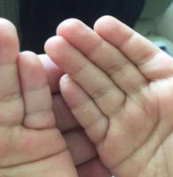
Skin exfoliation on the palms and soles of the feet for three days. He was initially seen and treated by a pediatrician in a private clinic, who prescribed an oral antibiotic to treat his symptoms. The child used the antibiotic without any improvement; he was then sent to the Pediatric Clinic of the National Guard Comprehensive Specialized Clinic in Riyadh, Saudi Arabia for further evaluation. His physical examination revealed fever with skin exfoliation on his palms and the soles of his feet (figure-1), significant tender lymphadenopathy over the posterior triangle of the left side of the neck, a strawberry tongue with cracked lips, and non-purulent conjunctivitis in both eyes. No skin rash was observed.
Investigation showed a total WBC count of 15.2 x10⁹ cells/L with 22% neutrophils, ESR (118 mm at the end of 1 hour), positive C-reactive protein, and high platelet count (1106 x 109/L). His anti-streptolysin-O titers were normal. The test for anti-nuclear antibody was negative. The throat swab culture was sterile. Liver enzymes were normal. Routine examination and culture of urine was negative. Echocardiograph was also normal. He was started on aspirin (100 mg/kg/day). His symptoms subsided within 24 hours of starting aspirin. He was discharged and maintained the same dosage of aspirin for one week, then the dose was reduced to (5 mg/kg/day) for the next 6 weeks. During his one week follow up, he was asymptomatic except for the exfoliation over on his hands and feet. Repeated CBC was markedly improved with WBC count 7.8 x10⁹ cells/L, and platelet count (543 x 109/L). ESR was 26. Two additional echocardiographs, one after 2 weeks and another after 6 weeks, were normal.
Case Report
A three-year-old Saudi male child presented with a history of high grade fever for 13 days, swelling in neck for seven days, and skin exfoliation on the palms and soles of the feet for three days. He was initially seen and treated by a pediatrician in a private clinic, who prescribed an oral antibiotic to treat his symptoms. The child used the antibiotic without any improvement; he was then sent to the Pediatric Clinic of the National Guard Comprehensive Specialized Clinic in Riyadh, Saudi Arabia for further evaluation. His physical examination revealed fever with skin exfoliation on his palms and the soles of his feet (figure-1), significant tender lymphadenopathy over the posterior triangle of the left side of the neck, a strawberry tongue with cracked lips, and non-purulent conjunctivitis in both eyes. No skin rash was observed. Investigation showed a total WBC count of 15.2 x10⁹ cells/L with 22% neutrophils, ESR (118 mm at the end of 1 hour), positive C-reactive protein, and high platelet count (1106 x 109/L). His anti-streptolysin-O titers were normal. The test for anti-nuclear antibody was negative. The throat swab culture was sterile. Liver enzymes were normal. Routine examination and culture of urine was negative. Echocardiograph was also normal. He was started on aspirin (100 mg/kg/day). His symptoms subsided within 24 hours of starting aspirin. He was discharged and maintained the same dosage of aspirin for one week, then the dose was reduced to (5 mg/kg/day) for the next 6 weeks. During his one week follow up, he was asymptomatic except for the exfoliation over on his hands and feet. Repeated CBC was markedly improved with WBC count 7.8 x10⁹ cells/L, and platelet count (543 x 109/L). ESR was 26. Two additional echocardiographs, one after 2 weeks and another after 6 weeks, were normal.
Figure-1: Exfoliation on the palms
Discussion
Kawasaki disease is the second most common cause of vasculitis in children after Henoch Schonlein purpura.[2,3] Early diagnosis and treatment of Kawasaki disease is of the utmost importance because of the dreadful complications that can occur during the acute illness. These complications include coronary arteritis, myocarditis, pericarditis, congestive heart failure and sudden death. The incidence of coronary aneurysm is around 20% of cases if left untreated. The diagnosis of Kawasaki disease is basically clinical and it is a diagnosis of exclusion. There are diagnostic criteria for the diagnosis of Kawasaki disease. For my case, I used the Japanese worker’s criteria.7 This patient had 4 criteria for the diagnosis of Kawasaki disease. Diagnostic criteria for kawasaki disease includes fever lasting for at least 5 days along with presence of at least 4 of the principal features: (1) Bilateral conjunctival injection, generally non-purulent, (2) Changes in the mucosa of the oropharynx, including injected oropharynx and dry fissured lips, and strawberry tongue, (3) Changes in the peripheral extremities such as edema and/or erythema of hands or feet during the acute phase, (4) Rash, primarily truncal, polymorphous, but non-vesicular, (5) Cervical lymphadenopathy (more than or equal to 1.5 cm in its diameter), usually unilateral. In addition to these criteria, illness should not be explained by other known disease processes.
In Kawasaki disease, it is very important to start treatment early, as this will help to reduce the risk of complications. The drug of choice in such situations will be a single dose of IVIG (2 g/kg) and then aspirin (100 mg/kg/day) for 14 days, followed by 3–5 mg/kg/day for 6 weeks.[8]
Treatment with IVIG relieves the acute inflammation and has been shown to reduce the rate of coronary aneurysms from more than 25% in untreated patients to 1-5% in treated patients. Maximum benefits are seen when IVIG is given within the first 10 days of the illness. Anti-inflammatory high dose aspirin (80-100 mg/kg/day orally divided into 4 doses) is given during the acute phase. Such dose should be continued until day 14 of the illness or until the patient has been afebrile for 48-72 hours, then a low-dose aspirin (3-5 mg/kg/day) is initiated for its antiplatelet activity for a total of 6-8 weeks provided patient shows no evidence of coronary abnormalities.[9,10] With prompt treatment, the prognosis of Kawasaki disease is good. The average mortality rate in the United States is approximately 1% of affected children. In patients younger than 1 year of age, the mortality rate may exceed 4%. In patients aged 1 year or older, the death rate is probably less than 1%. The mortality rate in japan is twice among boys with Kawasaki disease.
Conclusion
Kawasaki disease is terrifying vasculitis in a child. Any child presented with fever lasting more than a week, skin rash, peripheral extremities changes with edema or exfoliation, cracked lips, non-purulent conjunctivitis and cervical lymphadenopathy, should be investigated for Kawasaki disease. Prompt diagnosis and early institution of intravenous immunoglobulin and aspirin are of utmost importance in preventing coronary complications.
References
1. Laupland KB, Dele Davis H. Epidemiology, etiology, and management of Kawasaki disease: state of art. Pediatr Cardiol. 1999;20:177–83.
2. Melish ME, Hicks RV. Kawasaki syndrome: clinical features, pathophysiology, etiology and therapy. J Rheumatol Suppl. 1990;24:2-10.
3. Dhillon R, Newton L, Rudd PT, Hall SM. Management of Kawasaki disease in the British Isles. Arch Dis Child. 1993;69:631-8.
4.Taubert KA, Shulman ST. Kawasaki disease. Am Fam Physician. 1999;59:3093–102.
5.Aswine K Bal, Steven W Kairys. Kawasaki disease following Rocky Mountain spotted fever: a case report. Journal of Medical Case Reports. 2009;3:7320.
6.Sundel R.Clinical manifestations and diagnosis of Kawasaki disease [monograph on the Internet]. Waltham (MA): UpToDate, Inc; 2008 Jun 3. [cited 2008 Nov 4].
7.Japan Kawasaki Disease Research Committee. Diagnostic guidelines of Kawasaki disease, 4th rev. ed. Tokyo: Kawasaki Disease Research Committee; 1984.
8.Furusho K, Sato K, Soeda T, et al. High dose intravenous gammaglobulin for Kawasaki disease [letter]. Lancet. 1983;2:1359.
9.Newburger J W. Diagnosis, Treatment, and Long-Term Management of Kawasaki Disease: A Statement for Health Professionals From the Committee on Rheumatic Fever, Endocarditis, and Kawasaki Disease, Council on Cardiovascular Disease in the Young, American Heart Association Pediatrics. 2004;114;1708-1733.
10.Son M B F, Newburger J W. Management of Kawasaki disease: Corticosteroids Revisited. The lancet. 2012; 379:1571-1572.