4 Months Old Girl with Congenital Hypopigmentation (vitiligo) and Negative Family History of Vitiligo Born in Hayaat National Hospital-Riyadh (Saudi Arabia).
4 Months Old Girl with Congenital Hypopigmentation (vitiligo) and Negative Family History of Vitiligo Born in Hayaat National Hospital-Riyadh (Saudi Arabia).
Dr. Ahmed Moosa Assery*
*Correspondence to: Dr. Ahmed Moosa Assery. Paediatrics Ambulatory Care Consultant, KingAbdulaziz Medical City, National Guard Medical Affairs, Family Medicine-Ambulatory care, NGCSC.
Copyright
© 2017: Dr. Ahmed Moosa Assery. This is an open access article distributed under the Creative Commons Attribution License, which permits unrestricted use, distribution, and reproduction in any medium, provided the original work is properly cited.
Received: 10 September 2017
Published: 22 December 2017
Abstract
I will be proud to present such interesting case of an infant girl who born with diffuse hypopigmented spots in her body appeared from the first day of life that confused the treating medical staff in our institution (HNH) at that day including the paediatrics dermatologist who mentioned personally to me by this words: despite my long term experience and medical trip that took 35 years ,this the first time I face such dermatological presentation. The surprising thing in this case is the occurrence of this hypopigmentation spots (that labelled as a vitiligo later) which appeared in the body of the newborn soon after she got out to the life this because most of the references talking about the vitiligo mentioned the low rate of its prevalence as a congenital one. Not only this thing, the broad spectrum of the dermatological references which talked about the paediatrics vitiligo they mentioned the importance of the presence of the family history of vitiligo as a predisposing factor or at least presence of the any element of autoimmune disorder in the patient himself.
Keywords:
Hypopigmentation: HPOG.
Vitiligo: VG.
HNH: Hayaat National Hospital.
KSA: Kingdom of Saudi Arabia.
Riyadh: RDH.
Auto-immune Disorders: AID/s.
Juvenile Rheumatoid Arthritis: JRA
Systemic Lupus Erythematosus: SLE.
Thyroid Stimulating Hormone: TSH.
Phenyl Ketonuria: PKU.
Auditory Brainstem Response: ABR.
Ultra-violet: UV.
Anti-nuclear Antibodies: ANA.
Complete Blood Count: CBC.
Sedimentation Rate: ESR.
4 Months Old Girl with Congenital Hypopigmentation (vitiligo) and Negative Family History of Vitiligo Born in Hayaat National Hospital-Riyadh (Saudi Arabia).
Introduction
Vitiligo (VG) is a benign chronic HPOG disorder of the skin. It has a prevalence rate of 1 to 2 percent in the general population, with no predilection by sex, race, or region. The exact prevalence in the paediatric population is unknown.
Although aetiology of the disease is unknown, there are several theories including autoimmune and neurologic mechanisms, genetic factors, and the role of oxidative stress or toxic metabolites and lack of melanocyte growth factor. There are a few studies that assess congenital forms. However, the existence of these. Congenital forms are still controversial (1).
VG usually manifests in the second or third decade of life and is believed to be an acquired condition in almost half of the patients, it starts before 18 years of age and congenital forms are usually uncommon., though a positive family history is present in 30 to 40 percent of cases. Congenital VG and presentation at birth is a very rare entity, but cases in infancy have been reported one of them is our case here in this report. Due to its rare occurrence, there is a lack of data on congenital VG, and little is known about its exact etiopathogenesis and evolution. The known psychosocial impact of VG warrants more vigorous research into this disorder and increasing the scant knowledge with its respect (2).
In the old paediatrics patient’s occurrence of the VG (with different percentage number) can be associated with another AIDs like thyroiditis, pernicious anaemia, JRA, Type one IDDM T, SLE and psoriasis. That is why it is preferred for any new paediatrics VG case to do the investigations rolling out such disorders.
A survey in North America and the United Kingdom found that approximately 20 percent of 2624 vitiligo probands had a history of autoimmune thyroid disease compared with 2 percent of the general population (3).
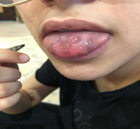
You can see here in this table in (Picture 1) how percent is the association of VG with other AIDs (4) in one study achieved in One hundred and 90 six consecutive adults’ patients with vitiligo referred to the Dermatology Department, University of La Laguna from September 2003 to September 2007.
Picture 1: More frequently associated diseases in our vitiligo patients
Case Presentation
This girl was born in June of 2017 in KSA-RDH in the secondary medical institution of HNH that is unfortunately lack of a research center, with normal non-eventful pre- and peri-natal history with full term pregnancy that passed without any complications. The delivery room medical staff who supervised the delivery (including the neonatologist) surprised about the white patches that girl born with in most of her body.
The newborn undergone the immediate routine post-natal blood screening (TSH – PKU – Bilirubin level) that all came normal then proceeded for the ABR which was also negative.
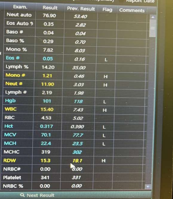
The paediatrics dermatologist involved in the second day in the inpatient setup and did an interview with the mother who denied any family history of VG or skin HPOG then the newborn undergone the UV Wood-lamp machine that used for the clinical assessment of the VG that came positive. (Picture 2):
The patient kept in the nursery 3 days more just for observation and no treatment started after which the newborn discharged with a follow up in after 6 months in both paediatrics and dermatology clinic.
When the infant became 6 months old, she presented to the combined paediatrics and dermatology clinic where the dermatologist recommended the start of the topical hydrocortisone creams and from the paediatrics side of view, I had done for the child some blood works that at least seek about any AIDs hose blood works were as follow:
*CBC+diff.
*Thyroids antibodies.
*ANA.
*Rheumatoid factor.
*ERS.
*C Reactive protein.
*Faecal occult blood.
*C3, C4 Complement components.
*Vitamin B12 level.
*Parietal cell antibodies.
The reason of selecting this age (6 months) to do such blood works for this child as known physiologically due to the just established real immune system of the infants that starts in this age in contrast to the earlier months in which the infants having only an artificial immunity taken from their mothers when they were fetuses. And, to pass the physiological anemia most of the infant got in the first 6 months of Age.
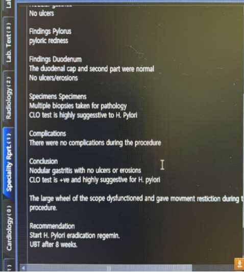
What was surprising is that all the results as in (Picture 3) came normal except with very high ANA reading. We tried to involve a paediatrics rheumatologist to take his/her opinion about that high ANA, but we could not.
The patient discharged with further follow up the combined treating team but unfortunately, they did not come back to search about will health future of the child.
Picture 2: this is the child in our case report with very wide and clear skin hypopigmented area in the abdomen
Picture 3: The laboratory blood test that showed here a very high ANA value
Discussion about the case
The interesting thing in this case is confined in two aspects first is being very rare to have a congenital VG, the second thing is the high ANA reading found in the blood works though the age of the child (6 months old) and the chance of AIDs associations in the future.
Regarding to being very seldom to have a congenital VG I just passed via the Medline search to collect as much as I can the reported cases that titled under the diagnosis of congenital VG and collected such cases:
A reported case published in Barro et al., 2017Licensee PAGEPress, ItalyPediatric Reports 2017; 9:7300doi:10.4081/pr.2017.7300. by Makoura Barro,1 Jean W. Diallo,2Ad Bafa Ibrahim Ouattara,1Boubacar Nacro11Department of Pediatrics, and2Department of Ophthalmology, SouroSanou Teaching Hospital, Bobo-Dioulasso, Burkina Faso that titled as Congenital vitiligo: A case.
observed in the cohort of HIV exposed infants in Bobo-Dioulasso, Burkina Faso that articled concluded that: Vitiligo at birth is very rare. Many other conditions can be involved and justify a structured diagnostic approach. Psychological support is insisted for such child when grown, the article said (5).
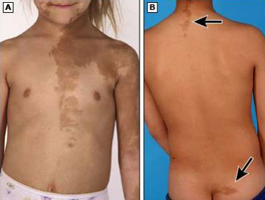
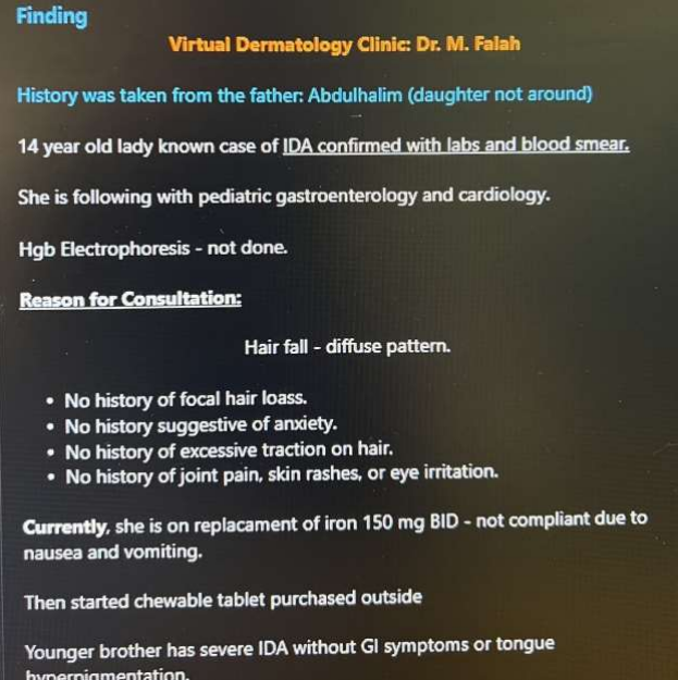
Another case reported for a 27- days-old female neonate patient presented with multiple, rapidly progressing, depigmented patches over the body that had been present since birth (Picture 4) that was in J Clin Aesthetic Dermatology. 2021 Jun; 14(6 Suppl 1): S27–S29.Published online 2021 Jun 1 by
Purva Pande, MD, Sree Ramu Suggu, MD, and Mala Bhalla, MD who concluded that Manifestation of VG at birth is a very rare occurrence. The presentation at birth in this case suggests a genetic link, as opposed to acquired factors, and supports the in-utero hypothesis, adding to the scant literature available on congenital VG (6).
Picture 4: Wood’s lamp examination showing chalky-white accentuation of lesions
Regarding to the possibility of the co-existing AIDs with congenital VG the searches taken about that are very rare (if not unavailable) I found here this article that was mainly established in adults and some paediatrics population that said:
Generalized VG is an AID in which acquired white patches of skin and overlying hair result from autoimmune loss of melanocytes from involved areas. Although usually sporadic, family clustering of VG may occur, in a non-Mendelian pattern typical of multifactorial, polygenic inheritance. Sporadic VG is associated with autoimmune thyroid disease, pernicious anemia, Addison's disease, and lupus; these same disorders occur at increased frequency in patients’ first-degree relatives. Here, we studied 133 ‘multiplex’ generalized VG families, with multiple affected family miembers. The age of onset of VG is earlier in these ‘multiplex’ families than in patients with sporadic VG. Affected members of the multiplex VG families have elevated frequencies of autoimmune thyroid disease, rheumatoid arthritis, psoriasis, adult-onset insulin-dependent diabetes mellitus, pernicious anemia, and Addison's disease. (7).
Conclusion
Though the prevalence of the congenital VG is very low, but it is very crucial to seek for the possible futural co-existing AIDS beginning from the 6 months old relaying on the family history and laboratory blood works.
References
11-https://www.ncbi.nlm.nih.gov/pmc/articles/PMC8565876/
2-https://www.ncbi.nlm.nih.gov/pmc/articles/PMC5573094/
3_https://www.uptodate.com/contents/vitiligo-pathogenesis-clinical-features-and- diagnosis? Source=history widget
4_https://www.researchgate.net/figure/More-frequently-associated-diseases-in-our-vitiligo-patients-n-196_tbl1_230624616
5_https://www.mdpi.com/2036-7503/9/3/7300
6_https://www.ncbi.nlm.nih.gov/pmc/articles/PMC8565876/#:~:text=Manifestation%20of%20vitiligo%20at%20birth,literature%20available%20on%20congenital%20vitiligo.
7_https://doi.org/10.1111/j.1600-0749.2005.00242.x