Serous Cystadenoma Torsion during Pregnancy in Multiparous A Case Report
Serous Cystadenoma Torsion during Pregnancy in Multiparous A Case Report
Acha Assmanni Adam*1, Rania E Belal2, Salma Mohammed Hassan Eltayeb3
1. Consultant of obstetrics and Gynecology in Saudia Arabia, MD Obstetrics and Gynecology. Medical Council of Sudan, Membership of Royal College of Obstetricians & Gynaecologists-UK.
2. Specialist of obstetrics and Gynecology in Saudia Arabia, MD Obstetrics and Gynecology, Medical Council of Sudan, Membership of Royal College of Obstetricians & Gynaecologists-UK.
3. OBGYN specialist /Tawam hospital –UAE.
*Correspondence to: Dr. Acha Bachir Assmanni Adam, Consultant of obstetrics and Gynecology in Saudia Arabia, MD Obstetrics and Gynecology. Medical Council of Sudan, Membership of Royal College of Obstetricians & Gynaecologists-UK.
Copyright
© 2024 Dr. Acha Bachir Assmanni Adam. This is an open access article distributed under the Creative Commons Attribution License, which permits unrestricted use, distribution, and reproduction in any medium, provided the original work is properly cited.
Received: 19 October 2024
Published: 23 October 2024
DOI: https://doi.org/10.5281/zenodo.13981136
Abstract
Ovarian cysts are common during pregnancy most cases are benign, but complications, such as torsion, and rupture can occur. The risk of torsion risk increases fivefold during pregnancy. It is associated with increased maternal and fetal morbidity and mortality if left untreated.
In Madina National Hospital, we report the case of A 36-year-old multiparous at 9 weeks, she has four previous cesarean section deliveries, the last baby was two years ago, presented to the emergency severe left iliac fossa with no radiating pain with nausea, and vomiting for 4 hours. was not radiated. Ultrasound diagnosed a left ovarian cyst. The patient was prepared for immediate laparotomy after resuscitation.
Intraoperative a cyst about 8cm with no signs of necrosis so detorsion and ovarian cystectomy were done, the cyst was removed intact, with minimal manipulation, and no intraoperative or postoperative complication, the other ovary was found to be normal. Histopathological examination revealed the presence of an ovarian serous cyst adenoma. She offered regular antenatal follow-up at our hospital with a healthy intrauterine fetus growing within.
Keywords: Ovarian, torsion, serous, cystadenoma, pregnancy.
Serous Cystadenoma Torsion during Pregnancy in Multiparous A Case Report
Introduction
Adnexal masses in pregnant women are 0.19-8.8%. The most common types are mature teratomas and para-ovarian or corpus luteum cysts. Ovarian masses during pregnancy can be functional, benign, or malignant neoplasms [1]. Serous cystadenoma rarely presents with ovarian cyst accidents in the first trimester as ovarian cysts appearing during the first trimester usually regress in size by the second trimester.[2]. During pregnancy ovarian cysts may cause pain and may cause ovarian torsion, requiring emergency management. Hence, women need an accurate assessment of their condition. Ovarian torsion commonly seen in the first trimester leads to necrosis which if untreated results in permanent loss of the ovary [3-4]. A common presentation of ovarian torsion is abdominopelvic pain, nausea, vomiting, and fever [5].
Case Presentation
A 36-year-old multiparous at 9 weeks, she has four previous cesarean section deliveries, last baby was two years ago, presented to the emergency severe left iliac fossa with no radiating pain with nausea, and vomiting for 4 hours. was not radiated.
Her symptoms are not relieved by simple analgesia; she has neither genitourinary symptoms nor gastrointestinal symptoms.
The patient gets spontaneous pregnancy, has no fever, she has no any medical illness. The patient had one visit at eight weeks of gestation and had no relevant medical or surgical history.
On examination, the patient looked unwell BP was 90/60 mmHg pulse rate was 114/min, and not febrile, cardiovascular, and respiratory systems were normal and the abdomen was rigid with rebound tenderness. A palpable mass at the left iliac fossa was noticed, vaginal examination showed fullness an urgent ultrasound was requested which showed a report attached, All of her blood and urine test results were within normal limits. Ultrasonography revealed normal filling of the urinary bladder with no stones or masses inside it.
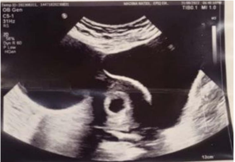
Anteverted gravid uterus, shows a single healthy pregnancy sac, with a healthy decidual reaction, it contains a single viable focal node with regular cardiac pulsations. RI 18mm-8 weeks 3days+/- 7 days Figure (1).
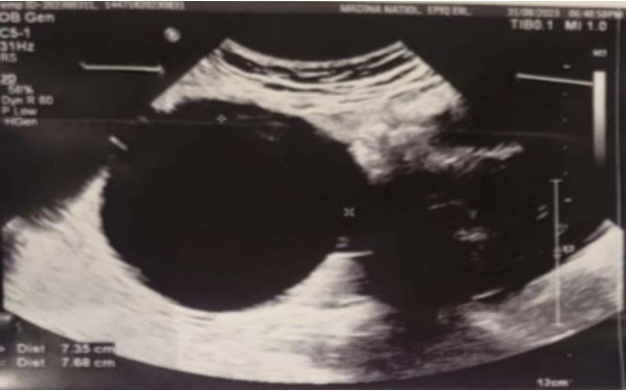
The left adnexa show a well-defined cystic lesion of about 6.7x8.6 cm, likely a left simple ovarian cyst, the cyst is surrounded by localized minimal to mild fluid collection around it, with tenderness on probing, by Doppler study, and it shows vascular flow however the possibility of ovarian torsion couldn't be excluded. Figure (2).
The right ovary is sonographically free with no solid or cystic lesions seen,with informed consent given for an acute emergency, the patient was shifted for emergency laparotomy under spinal anesthesia, intraoperative some adhesions were dissected, and the ovary was tortured 3 times with a cyst about 8cm with no signs of necrosis so detorsion and ovarian cystectomy were done, the cyst was removed intact, with minimal manipulation, and no intraoperative or postoperative complication, the other ovary was found to be normal.
Post-operative care; The patient received analgesia and rehydration according to the hospital protocol along with progesterone suppository for up to 16 weeks.
She was discharged in good condition after 48 hours. A normal postoperative obstetric ultrasound suggesting a live 9-week fetus was observed on the third postoperative.
Histology result reports ovarian cyst with smooth surface measuring 9.5x9x6 con. The thickness of the wall measuring 0.1-0.5 cm, shows a discoloration measuring counter 3.5 cm.
Microscopy showed the excised tissue shows ovarian tissue with serous cystadenoma lined by containing corpus luteum, negative for e for borderline lesion or malignancy. flat and cuboidal epithelium with fibrous wall.
Final diagnosis Serous cystadenoma -Negative for borderline lesion or malignancy.
Fig 1, Fig 2
Discussion
Serous cystadenomas, luteomas, corpus luteal cysts, and cystic teratomas are most common the ovarian tumors presented during pregnancy [6]. Ovarian serous cystadenomas are common benign epithelial neoplasms with an excellent prognosis, representing 16% of all ovarian epithelial neoplasms, occurring in adults of all ages and bilateral in 10 to 20% of women [7]. Serous cystadenoma can be found in ultrasound it is a size from one centimeter to more than thirty cm in greatest dimension it is usually unilocular but it can be multilocular [8].
Serous cystadenomas are composed of cysts and papillae lined by non-stratified or stratified cuboidal to columnar cells like fallopian tube epithelium.[9]. The immunohistochemical profile of serous cystadenoma is similar to that of normal ovarian surface epithelium and tubal epithelium. In addition to positivity with most commonly used epithelial markers, p63 is positive in most cases
[10] .
The measurement of serum CA-125 assay is a useful tool that helps distinguish between benign and malignant ovarian masses. The combination of normal findings at serum CA-125 assay, imaging, and clinical presentation findings exclude the possibility of ovarian cancer [11].
Serous cystadenoma has an excellent prognosis with a risk of recurrence after cystectomy [12]. Presentation may be asymptomatic, rare complications of ovarian cystadenomas include ovarian torsion or cyst rupture [13]. Ovarian torsion commonly occurs in the first trimester and is least during the third trimester [14].
Although emergency exploratory laparotomy may be necessary to treat twisting or infarction and rupture ovarian cysts, regardless of the stage of pregnancy, an ovarian cyst that ruptures, or twists must be surgically treatment to reduce the risk of complications in both the mother and fetus [15].
The availability of transvaginal color Doppler, MRI, and tumor markers improves the management of ovarian accidents in pregnancy.
Conclusions
Serous cystadenoma ovarian torsion during pregnancy is rare and presentation with torsion in early gestation as ovarian torsions since only 1%-2% of pregnant women present with this condition and most cases cannot be detected. If the cyst is twisted or ruptured it can lead to significant morbidity. The obstetrician should be aware of the possibility of ovarian torsion in pregnant women and increase the index of suspicion. With the aid of ultrasound Doppler and tumor markers and surgical intervention both maternal and fetal morbidity will be reduced and good obstetrics outcomes will be obtained.
Conflicts of interest: No conflict of interest
Financial relationships: Not funded
Other relationships: No other relationships or activities that could appear to have influenced the submitted work.
References
1. Cavaco-Gomes J, Jorge Moreira C, Rocha A, Mota R, Paiva V, Costa A. Investigation and Management of Adnexal Masses in Pregnancy. Scientifica (Cairo). 2016;2016:3012802.
2. Cunningham FG, Leveno KJ, Bloom SL, Hauth JC, Rouse DJ, Spong CY. Reproductive tract abnormalities. 23rd ed. New York: McGraw-Hill; 2010:912-253.
3. Feng J-L, Zheng J, Lei T, Xu Y-J, Pang H, Xie H-N. Comparison of ovarian torsion between pregnant and non-pregnant women at reproductive ages: Sonographic and pathologic findings. Quant Imaging Med Surg. 2020;10(1):137−47.
4.Young R, Cork K. Intermittent ovarian torsion in pregnancy. Clin Pract Cases Emerg Med. 2017;1(2):108−10. doi:10.5811/cpcem.2016. 12.32932.
5. Lourenco AP, Swenson D, Tubbs RJ, Lazarus E. Ovarian and tubaltorsion: Imaging findings on US, CT, and MRI. Emerg Radiol 2014; 21:179-187
6. Bhagat, N., & Gajjar, K. (2022). Management of ovarian cysts during pregnancy.
Obstetrics, Gynaecology & Reproductive Medicine, 32(9), 205-210.
7. Ben?Mussa, A., & McCluggage, W. G. (2021). Ovarian seromucinous cystadenomas and adenofibromas: first report of a case series. Histopathology, 78(3), 445-452.
8. Shukla, N. (2020). Role of DWI in Differentiating Between Benign and Malignant Uterine and Adnexal Lesions (Doctoral dissertation, Rajiv Gandhi University of Health Sciences (India)).
9. Georgescu, T. A., Bohiltea, R., Munteanu, O., Grigoriu, C., Paunica, I., & Sajin, M. (2021). A mini-review regarding the carcinogenesis and morphology of serous tumors of the ovary, fallopian tube and peritoneum. Journal of Mind and Medical Sciences, 8(1), 44-52.
10. Poli Neto OB, Candido Dos Reis FJ, Zambelli Ramalho LN, Nogueira AA, de Andrade JM. P63 expression in epithelial ovarian tumors. Int J Gynecol Cancer. 2006 Jan- Feb;16(1):152-5. [PubMed]
11. Elorriaga, M. Á., Neyro, J. L., Mieza, J., Cristóbal, I., & Llueca, A. (2021). Biomarkers in ovarian pathology: from screening to diagnosis. Review of the literature. Journal of Personalized Medicine, 11(11), 1115.
12. Foula, M. S., AlQattan, A. S., AlQurashi, A. M., AlShaqaq, H. M., & Gari, M. K. M. (2019). Incidentally discovered huge retroperitoneal mucinous cystadenoma with successful laparoscopic management: case report. International journal of surgery case reports, 61, 242-245.
13. Grigore, M., Murarasu, M., Himiniuc, L. M., Toma, B. F., Duma, O., & Popovici, R. (2021). Large ovarian tumors in adolescents, a systematic review of reported cases, diagnostic findings and surgical management. Taiwanese Journal of Obstetrics and Gynecology, 60(4), 602-608.
14. Meller, N., Levin, G., Cohen, A., Abu?Bandora, E., Amitai Komem, D., Mashiach, R., & Meyer, R. (2021). Surgically confirmed adnexal torsion during pregnancy: Does the trimester make a difference?. Journal of Obstetrics and Gynaecology Research, 47(12), 4216-4223.
15. Spinelli, C., Tröbs, R. B., Nissen, M., Strambi, S., Ghionzoli, M., Bertocchini, A., ... & Lelli Chiesa, P. (2023). Ovarian torsion in the pediatric population: Predictive factors for ovarian-sparing surgery—An international retrospective multicenter study and a systematic review. Archives of Gynecology and Obstetrics, 308(1), 1-12..